

An Integrated System for Superharmonic Contrast-Enhanced Ultrasound Imaging: Design and Intravascular Phantom Imaging Study
- PMID: 26672030
- PMCID: PMC6535182
- DOI: 10.1109/TBME.2015.2506639
An Integrated System for Superharmonic Contrast-Enhanced Ultrasound Imaging: Design and Intravascular Phantom Imaging Study
Abstract
Objective: Superharmonic contrast-enhanced ultrasound imaging, also called acoustic angiography, has previously been used for the imaging of microvasculature. This approach excites microbubble contrast agents near their resonance frequency and receives echoes at nonoverlapping superharmonic bandwidths. No integrated system currently exists could fully support this application. To fulfill this need, an integrated dual-channel transmit/receive system for superharmonic imaging was designed, built, and characterized experimentally.
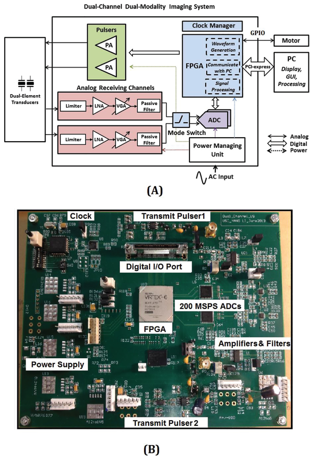
Method: The system was uniquely designed for superharmonic imaging and high-resolution B-mode imaging. A complete ultrasound system including a pulse generator, a data acquisition unit, and a signal processing unit were integrated into a single package. The system was controlled by a field-programmable gate array, on which multiple user-defined modes were implemented. A 6-, 35-MHz dual-frequency dual-element intravascular ultrasound transducer was designed and used for imaging.
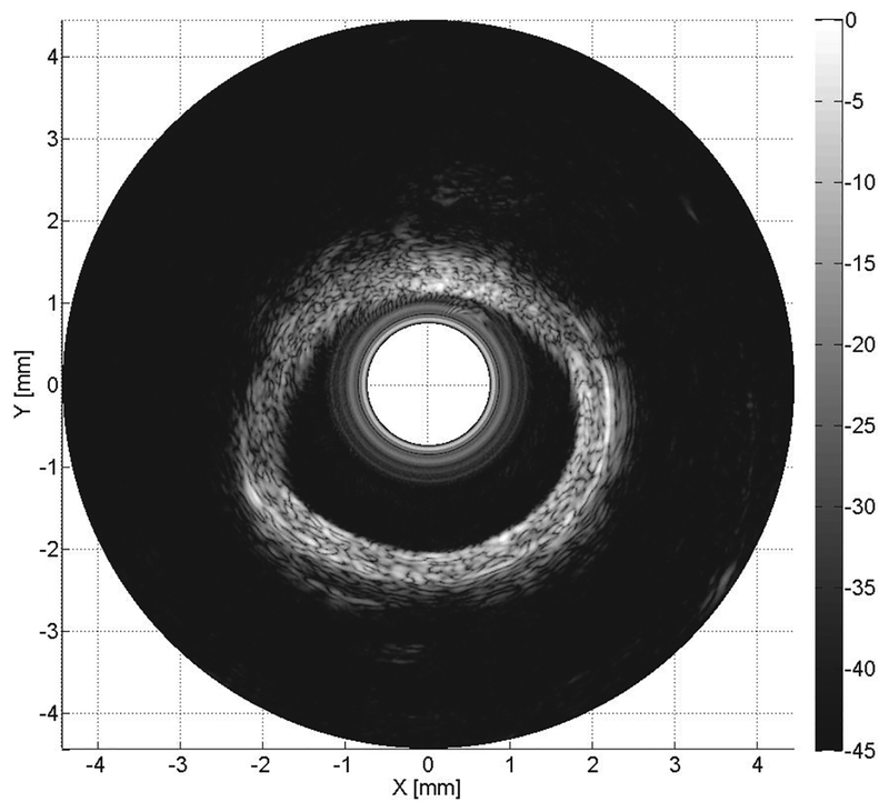
Result: The system successfully obtained high-resolution B-mode images of coronary artery ex vivo with 45-dB dynamic range. The system was capable of acquiring in vitro superharmonic images of a vasa vasorum mimicking phantom with 30-dB contrast. It could detect a contrast agent filled tissue mimicking tube of 200 μm diameter.
Conclusion: For the first time, high-resolution B-mode images and superharmonic images were obtained in an intravascular phantom, made possible by the dedicated integrated system proposed. The system greatly reduced the cost and complexity of the superharmonic imaging intended for preclinical study. Significant: The system showed promise for high-contrast intravascular microvascular imaging, which may have significant importance in assessment of the vasa vasorum associated with atherosclerotic plaques.
Figures


Similar articles
-
Molecular Acoustic Angiography: A New Technique for High-resolution Superharmonic Ultrasound Molecular Imaging.Ultrasound Med Biol. 2016 Mar;42(3):769-81. doi: 10.1016/j.ultrasmedbio.2015.10.015. Epub 2015 Dec 8. Ultrasound Med Biol. 2016. PMID: 26678155 Free PMC article.
-
Adaptive windowing in contrast-enhanced intravascular ultrasound imaging.Ultrasonics. 2016 Aug;70:123-35. doi: 10.1016/j.ultras.2016.04.022. Epub 2016 Apr 27. Ultrasonics. 2016. PMID: 27161022 Free PMC article.
-
Ex Vivo Porcine Arterial and Chorioallantoic Membrane Acoustic Angiography Using Dual-Frequency Intravascular Ultrasound Probes.Ultrasound Med Biol. 2016 Sep;42(9):2294-307. doi: 10.1016/j.ultrasmedbio.2016.04.008. Epub 2016 May 31. Ultrasound Med Biol. 2016. PMID: 27260246 Free PMC article.
-
Emerging technology in head and neck ultrasonography.Otolaryngol Clin North Am. 2010 Dec;43(6):1267-74, vii. doi: 10.1016/j.otc.2010.08.003. Otolaryngol Clin North Am. 2010. PMID: 21044741 Review.
-
Visualization of Microvascular Angiogenesis Using Dual-Frequency Contrast-Enhanced Acoustic Angiography: A Review.Ultrasound Med Biol. 2020 Oct;46(10):2625-2635. doi: 10.1016/j.ultrasmedbio.2020.06.009. Epub 2020 Jul 20. Ultrasound Med Biol. 2020. PMID: 32703659 Free PMC article. Review.
Cited by
-
Recent Advances in Transducers for Intravascular Ultrasound (IVUS) Imaging.Sensors (Basel). 2021 May 19;21(10):3540. doi: 10.3390/s21103540. Sensors (Basel). 2021. PMID: 34069613 Free PMC article. Review.
-
Intravascular Ultrasound Imaging With Virtual Source Synthetic Aperture Focusing and Coherence Factor Weighting.IEEE Trans Med Imaging. 2017 Oct;36(10):2171-2178. doi: 10.1109/TMI.2017.2723479. Epub 2017 Jul 4. IEEE Trans Med Imaging. 2017. PMID: 28692968 Free PMC article.
-
Dual-Resonance (16/32 MHz) Piezoelectric Transducer With a Single Electrical Connection for Forward-Viewing Robotic Guidewire.IEEE Trans Ultrason Ferroelectr Freq Control. 2022 Apr;69(4):1428-1441. doi: 10.1109/TUFFC.2022.3150746. Epub 2022 Mar 30. IEEE Trans Ultrason Ferroelectr Freq Control. 2022. PMID: 35143395 Free PMC article.
References
-
- Alwan A, Global status report on noncommunicable diseases 2010: World Health Organization, 2011.
-
- Olsson O, “Vertebral Angiography in the Diagnosis of Acoustic Nerve Tumours,” Acta Radiologica, vol. 39, pp. 265–272, 1953. - PubMed
-
- Naghavi M, Libby P, Falk E, Casscells SW, Litovsky S, Rumberger J, et al., “From vulnerable plaque to vulnerable patient a call for new definitions and risk assessment strategies: part I,” Circulation, vol. 108, pp. 1664–1672, 2003. - PubMed
-
- Muller JE, Tofler GH, and Stone PH, “Circadian variation and triggers of onset of acute cardiovascular disease,” Circulation, vol. 79, pp. 733–43, April 1989. - PubMed
-
- Moreno PR, Purushothaman KR, Fuster V, Echeverri D, Truszczynska H, Sharma SK, et al., “Plaque neovascularization is increased in ruptured atherosclerotic lesions of human aorta: implications for plaque vulnerability,” Circulation, vol. 110, pp. 2032–8, October 5 2004. - PubMed
Publication types
MeSH terms
Substances
Grants and funding
LinkOut - more resources
Full Text Sources
Other Literature Sources
Miscellaneous
