Review
. 2015 Dec 15;142(24):4191-204.
doi: 10.1242/dev.114777.

Tendon development and musculoskeletal assembly: emerging roles for the extracellular matrix

Affiliations
Review

Tendon development and musculoskeletal assembly: emerging roles for the extracellular matrix

Arul Subramanian et al. Development. .

Abstract

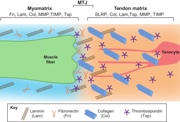
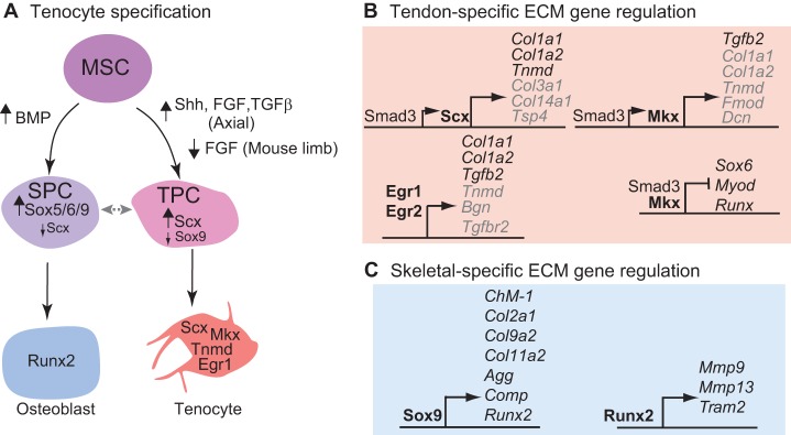
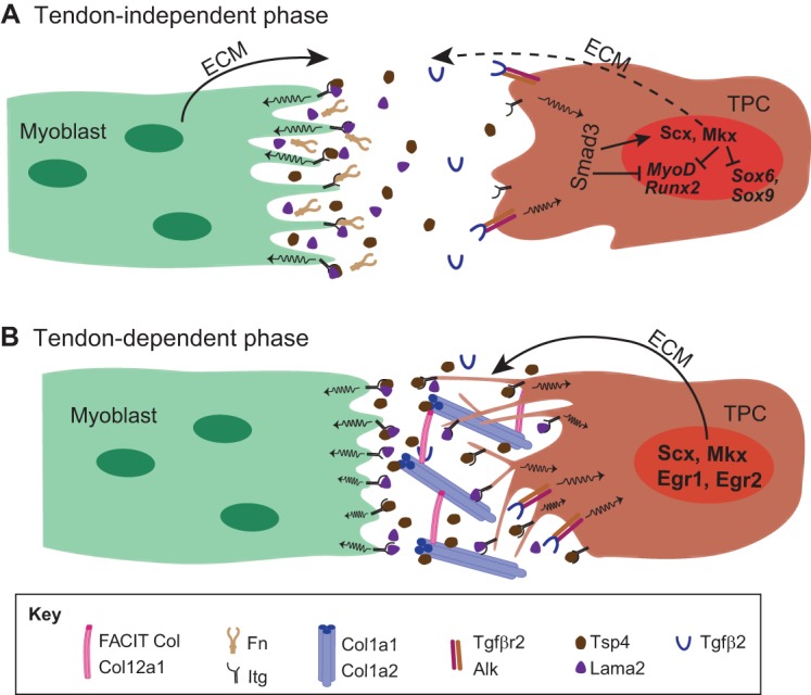
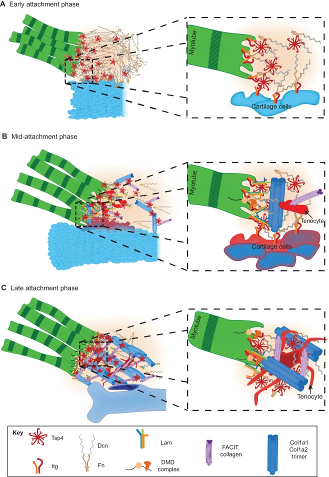
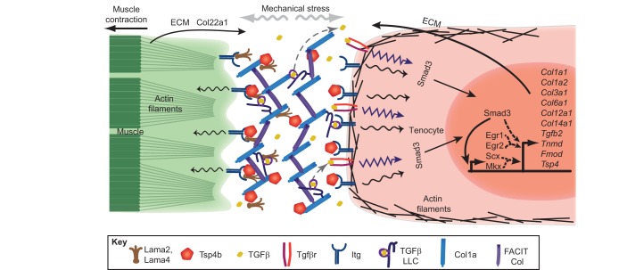
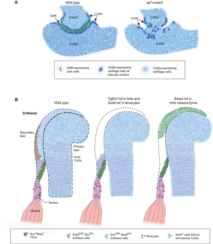
Tendons and ligaments are extracellular matrix (ECM)-rich structures that interconnect muscles and bones. Recent work has shown how tendon fibroblasts (tenocytes) interact with muscles via the ECM to establish connectivity and strengthen attachments under tension. Similarly, ECM-dependent interactions between tenocytes and cartilage/bone ensure that tendon-bone attachments form with the appropriate strength for the force required. Recent studies have also established a close lineal relationship between tenocytes and skeletal progenitors, highlighting the fact that defects in signals modulated by the ECM can alter the balance between these fates, as occurs in calcifying tendinopathies associated with aging. The dynamic fine-tuning of tendon ECM composition and assembly thus gives rise to the remarkable characteristics of this unique tissue type. Here, we provide an overview of the functions of the ECM in tendon formation and maturation that attempts to integrate findings from developmental genetics with those of matrix biology.

Keywords: Extracellular matrix; Ligament; Tendon; Tenocyte.


Conflict of interest statement

Competing interests

The authors declare no competing or financial interests.

Figures

Fig. 1.
Fig. 1.
Composition of the ECM surrounding muscle, tendon and myotendinous junctions. A muscle fiber (green) secretes ECM components into its surroundings (the myomatrix). Some of these components overlap with those of the tendon ECM, which is secreted by tenocytes (red). Myomatrix is primarily composed of Lam trimers and Fn. By contrast, the tendon matrix is rich in Col1a trimers and thrombospondin pentamers. The myotendinous junction (MTJ) is the narrow zone in which ECM components of tendon and muscle interact.
Fig. 2.
Fig. 2.
Transcriptional regulation of tenocyte specification and tendon ECM production. (A) A mesenchymal stem cell (MSC, purple), the common progenitor for skeletal and tenocyte progenitors, becomes a skeletogenic progenitor cell (SPC, blue) if it is exposed to high levels of BMP signaling, then it expresses Sox5/6/9 followed by Runx2 during its differentiation into an osteoblast. By contrast, an MSC becomes a tendon progenitor cell (TPC, pink) if it receives high levels of Shh, FGF and TGFβ signaling, then expresses scleraxis (Scx) followed by Mkx, Egr1 and Tnmd during its transition into a tenocyte. The fate of the progenitor cells is determined by the level of Sox9 and Scx. The plasticity of progenitor cell fate at this stage is represented by the double-headed gray arrow. (B) The transcription factors involved in tenocyte specification also regulate the transcription of genes encoding ECM proteins. Direct (black) and indirect (gray) transcriptional target genes regulated by Scx, Egr1, Egr2 and Mkx in TPCs are indicated. Note that Mkx also represses the expression of factors involved in myogenic and skeletogenic progenitor formation. (C) Transcription factors expressed in skeletogenic progenitor cells (e.g. Sox9 and Runx2) directly regulate the transcription of a distinct set of ECM target genes.
Fig. 3.
Fig. 3.
Myoblast-tenocyte interactions and ECM production. (A,B) The formation of myotendinous junctions can be considered as a two-step process. In the initial tendon-independent phase (A) in vertebrates (shown here for zebrafish trunk muscles), myoblasts (green) synthesize a ‘pre-tendon’ ECM that includes the integrin ligands Tsp4 and Lama2. This ECM accumulates in the absence of TPCs (brown). Mechanotransduction coupled with TGFβ signaling (through Tgfβ2 and Tgfβr2) leads to the Smad3-dependent expression of Scx and Mkx in TPCs, which in turn leads to the expression of tendon-selective ECM genes. Smad3 and Mkx also repress the activity of MyoD, Sox9 and Runx2 to repress myogenic and skeletogenic fates during tenocyte differentiation. A later tendon-dependent phase (B) relies on the production of ECM, particularly Col1a1, Col1a2, Col12a1 and Col14a1, by more mature TPCs, which extend processes into the ECM.
Fig. 4.
Fig. 4.
Maturation and assembly of the tendon ECM. Diagram illustrating progressive changes in the ECM at an MTJ as it matures. (A) In the early attachment phase, myoblasts (green) first extend towards a cartilage condensation (blue) and reorganize the local ECM by secreting Tsp4 (red), which interacts with Fn and Lam. A magnified view (right) of the boxed area illustrates how Tsp4 pentamers assemble Fn, Lam and Dcn and facilitate binding to Itgs on both muscle and cartilage cell surfaces, thereby promoting adhesion. (B) Following this, in the mid-attachment phase, linear collagen fibrils (Col1a1 trimers, dark blue) form, tenocytes (red) invade, and Sox9+/Scx+ progenitors (dark blue and orange) become detected at the future attachment site on the cartilage, the enthesis. The magnified view illustrates how Col1a1 trimers begin to align perpendicular to skeletal cells (enthesis, dark blue and orange). Dystrophin (DMD) complexes appear on muscle surfaces. (C) In the final late attachment phase, collagen fibrils become crosslinked into a lattice, with tenocytes (red cells) extending processes to surround fibrils, and entheses chondrifying (purple). The magnified view shows Col1a1 trimers becoming crosslinked by FACIT collagens and surrounded by tenocyte (red) processes, stabilizing the ECM and its interactions with Itgs on muscle and cartilage cells.
Fig. 5.
Fig. 5.
Model for ECM-mediated feedback from mechanical force and its effects on tenocyte gene expression. A tenocyte (orange) synthesizes the tendon ECM, including Lama2, Lama4 (brown), Tsp4b (red pentagons), Col1a (blue) and FACIT Col (purple), all of which signal through Itg receptors (dark blue) on muscle and tenocyte cell surfaces in response to mechanical stress (gray arrows). In addition, stress causes the ECM to release TGFβ (yellow) from the TGFβ large latent complex (LLC) (gray dotted arrows). Itg and TGFβ signaling in tenocytes feedback to regulate Scx-, Egr1/2- and Mkx-induced transcription (dashed arrows) of the same Itg ligands as well as of other ECM components to modulate tendon stiffness. Smad3 also interacts with Scx and Mkx to activate target ECM genes. The muscle fiber also contributes to the tendon matrix by secreting FACIT Col22a1.
Fig. 6.
Fig. 6.
ECM functions at tendon-bone attachments. (A) Diagrams illustrating changes in cartilage at the developing humero-ulnar joint of the mouse forelimb in wild-type embryos (left) and in splotch delayed (spd, Pax3) mutants (right), which lack muscles. Proliferating chondrocytes express Col2a1 (light blue), whereas cells forming at the edges of the joint express Col2b (dark blue), and cells in the joint interzone secrete Gdf5 (green) into the joint region. The loss of muscles in spd mutants leads to loss of Gdf5 expression, disorganized Col2b+ interzone cells and joint fusion. (B) Diagrams illustrating changes in cartilage and tenocytes at a developing eminence. The primary field contains cells that form chondrocytes within the developing bone, whereas the secondary field consists of Sox9-positive progenitor cells that lie outside of the primary field. In wild-type embryos, three different subsets of Scx-expressing cells at a muscle insertion site of a developing long bone are found: Sox9+/Scx+, Scx+ or Scx+/Bmp4+. Loss of Tgfβr2 in limb mesenchyme or of Sox9 in tenocytes leads to a loss of the Sox9/Scx co-expressing and Sox9-expressing population in the secondary field, but not other tenocytes. Loss of Bmp4 signaling leads to a loss of both Sox9+/Scx+ and Scx+ populations in the secondary field. Dotted lines outline primary field. Dashed lines outline secondary field. lof, loss of function.

Similar articles

Cited by

References

    1. Abzhanov A., Protas M., Grant B. R., Grant P. R. and Tabin C. J. (2004). Bmp4 and morphological variation of beaks in Darwin's finches. Science 305, 1462-1465. 10.1126/science.1098095 - DOI - PubMed
    1. Adams J. C. and Lawler J. (2004). The thrombospondins. Int. J. Biochem. Cell Biol. 36, 961-968. 10.1016/j.biocel.2004.01.004 - DOI - PMC - PubMed
    1. Alberton P., Popov C., Prägert M., Kohler J., Shukunami C., Schieker M. and Docheva D. (2012). Conversion of human bone marrow-derived mesenchymal stem cells into tendon progenitor cells by ectopic expression of scleraxis. Stem Cells Dev. 21, 846-858. 10.1089/scd.2011.0150 - DOI - PMC - PubMed
    1. Albertson R. C., Streelman J. T., Kocher T. D. and Yelick P. C. (2005). Integration and evolution of the cichlid mandible: the molecular basis of alternate feeding strategies. Proc. Natl. Acad. Sci. USA 102, 16287-16292. 10.1073/pnas.0506649102 - DOI - PMC - PubMed
    1. Alliston T., Choy L., Ducy P., Karsenty G. and Derynck R. (2001). TGF-beta-induced repression of CBFA1 by Smad3 decreases cbfa1 and osteocalcin expression and inhibits osteoblast differentiation. EMBO J. 20, 2254-2272. 10.1093/emboj/20.9.2254 - DOI - PMC - PubMed

Publication types

LinkOut - more resources