. 2016 Feb 2;7(5):5273-88.
doi: 10.18632/oncotarget.6567.

Unscrambling the genomic chaos of osteosarcoma reveals extensive transcript fusion, recurrent rearrangements and frequent novel TP53 aberrations

Affiliations

Unscrambling the genomic chaos of osteosarcoma reveals extensive transcript fusion, recurrent rearrangements and frequent novel TP53 aberrations

Susanne Lorenz et al. Oncotarget. .

Abstract

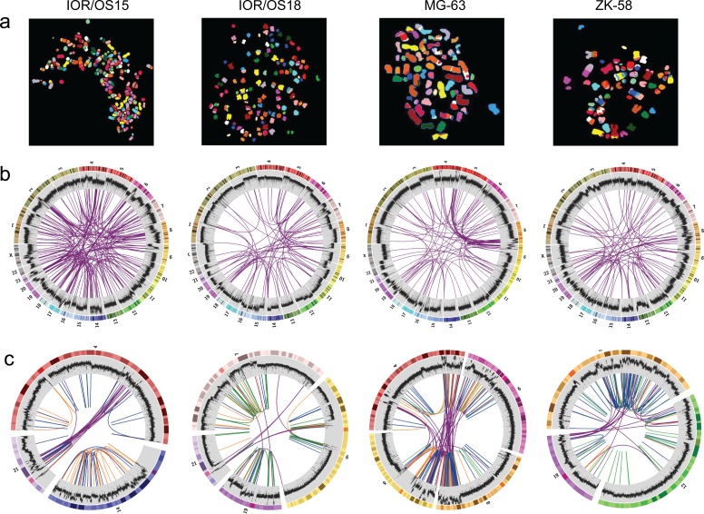
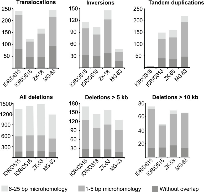
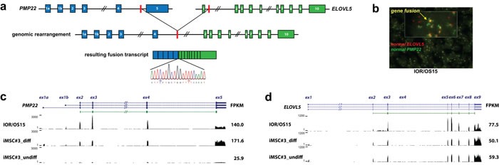
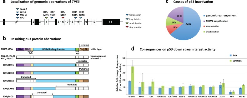
In contrast to many other sarcoma subtypes, the chaotic karyotypes of osteosarcoma have precluded the identification of pathognomonic translocations. We here report hundreds of genomic rearrangements in osteosarcoma cell lines, showing clear characteristics of microhomology-mediated break-induced replication (MMBIR) and end-joining repair (MMEJ) mechanisms. However, at RNA level, the majority of the fused transcripts did not correspond to genomic rearrangements, suggesting the involvement of trans-splicing, which was further supported by typical trans-splicing characteristics. By combining genomic and transcriptomic analysis, certain recurrent rearrangements were identified and further validated in patient biopsies, including a PMP22-ELOVL5 gene fusion, genomic structural variations affecting RB1, MTAP/CDKN2A and MDM2, and, most frequently, rearrangements involving TP53. Most cell lines (7/11) and a large fraction of tumor samples (10/25) showed TP53 rearrangements, in addition to somatic point mutations (6 patient samples, 1 cell line) and MDM2 amplifications (2 patient samples, 2 cell lines). The resulting inactivation of p53 was demonstrated by a deficiency of the radiation-induced DNA damage response. Thus, TP53 rearrangements are the major mechanism of p53 inactivation in osteosarcoma. Together with active MMBIR and MMEJ, this inactivation probably contributes to the exceptional chromosomal instability in these tumors. Although rampant rearrangements appear to be a phenotype of osteosarcomas, we demonstrate that among the huge number of probable passenger rearrangements, specific recurrent, possibly oncogenic, events are present. For the first time the genomic chaos of osteosarcoma is characterized so thoroughly and delivered new insights in mechanisms involved in osteosarcoma development and may contribute to new diagnostic and therapeutic strategies.

Keywords: DNA repair; bone cancer; gene fusion; osteosarcomas; trans-splicing.


Conflict of interest statement

CONFLICTS OF INTEREST

The authors disclose no potential conflicts of interest.

Figures

Figure 1
Figure 1. Visualization of the genomic chaos in osteosarcoma
a. Example single cell multicolor spectral karyotypes for each cell line demonstrating the genomic complexity and high numbers of translocations. b. Circos plots showing genome-wide interchromosomal translocations (purple lines) identified by WGS with allele frequencies of ≥10 %. The outermost circles illustrate the chromosome idiograms followed by the plot of the genome coverage (binary logarithmic scale ranging from 2 (log24) to 8 (log2256), increasing from center towards periphery, 50K window size). Changes in coverage indicate copy number variations with increase indicating gain and decrease indicating loss. c. Circos plots of rearrangement clusters showing chromothripsis-like characteristics of breakpoint distribution within certain chromosomes. Purple lines indicate translocations, orange lines inversions, green lines duplications and blue lines deletions. The coverage plot illustrates high local increase of the copy number, which does not support the copy number neutral chromothripsis model.
Figure 2
Figure 2. Abundance of the different genomic rearrangement types and their microhomology pattern
The rearrangements are grouped by type (translocations, inversions, tandem duplications, deletions) and divided in three categories of overlapping microhomology at the breakpoints: 6-25 bp overlap, 1-5 bp overlap and without overlap. Deletions are further divided by length, showing that smaller deletions (< 5 kb) have a different microhomology pattern with a higher frequency of 6-25 bp long overlapping microhomology. This frequency was clearly reduced for deletions longer than 5 kb and more similar to the other types of rearrangements. The same tendency was found for deletions longer than 10 kb, which showed even more similarity to the microhomology pattern of translocations, inversions and tandem duplications.
Figure 3
Figure 3. PMP22-ELOVL5 gene fusion
a. Schematic overview of the location of the genomic breakpoint and the resulting fusion transcript identified in the cell line IOR/OS15. Validation of the fusion transcript by Sanger sequencing confirmed the breakpoint as illustrated in the sequence chromatogram. b. Double-fusion FISH picture of IOR/OS15 in tissue microarray (cell pellets). Green and red probes identify PMP22 and ELOVL5, respectively. Colocalized signals are visible in yellow and confirm the gene fusion at the genome level. Further, normalized expression patterns of PMP22 c. and ELOVL5 d. are shown for IOR/OS15, differentiated and undifferentiated iMSC#3 with the corresponding FPKM (Fragments Per Kilobase of exon per Million fragments mapped) calculated using Cufflinks. For IOR/OS15 a reduced expression of PMP22 exon 5 and ELOVL5 exon 1 is visible, caused by the loss of these exons in the fusion transcript.
Figure 4
Figure 4. TP53 aberrations in osteosarcoma cell lines
a. Genomic localization of aberrations across TP53 with triangles demonstrating the position of the aberrations for the different cell lines and the aberration type (dark blue = translocation, light blue = long deletion, green = small deletion, red = stop mutation). b. Visualization of the consequences of the TP53 aberrations at the protein level. The different protein domains are indicated (TAD = transactivation domain, NLS = nuclear localization signal, OD = oligomerization domain, BR = basic region). Uncolored regions of the protein represent truncated parts. c. The distribution of the various causes of p53 inactivation clearly shows that rearrangements in TP53 are the major factor with 64 % followed by MDM2 amplifications, small deletions and mutations. d. Results of the radiation-induced DNA damage assay showing the fold change of BAX and CDKN1A expression 8 hours after induction. U-2 OS is known to harbor wild-type p53 and was used as control.

References

    1. Bayani J, Zielenska M, Pandita A, Al-Romaih K, Karaskova J, Harrison K, Bridge JA, Sorensen P, Thorner P, Squire JA. Spectral karyotyping identifies recurrent complex rearrangements of chromosomes 8, 17, and 20 in osteosarcomas. Genes Chromosomes Cancer. 2003;36:7–16. - PubMed
    1. Kuijjer ML, Rydbeck H, Kresse SH, Buddingh EP, Lid AB, Roelofs H, Burger H, Myklebost O, Hogendoorn PC, Meza-Zepeda LA, Cleton-Jansen AM. Identification of osteosarcoma driver genes by integrative analysis of copy number and gene expression data. Genes Chromosomes Cancer. 2012;51:696–706. - PubMed
    1. Kresse SH, Rydbeck H, Skarn M, Namlos HM, Barragan-Polania AH, Cleton-Jansen AM, Serra M, Liestol K, Hogendoorn PC, Hovig E, Myklebost O, Meza-Zepeda LA. Integrative analysis reveals relationships of genetic and epigenetic alterations in osteosarcoma. PLoS One. 2012;7:e48262. - PMC - PubMed
    1. Kresse SH, Ohnstad HO, Paulsen EB, Bjerkehagen B, Szuhai K, Serra M, Schaefer KL, Myklebost O, Meza-Zepeda LA. LSAMP, a novel candidate tumor suppressor gene in human osteosarcomas, identified by array comparative genomic hybridization. Genes Chromosomes Cancer. 2009;48:679–693. - PubMed
    1. Baroy T, Kresse SH, Skarn M, Stabell M, Castro R, Lauvrak S, Llombart-Bosch A, Myklebost O, Meza-Zepeda LA. Reexpression of LSAMP inhibits tumor growth in a preclinical osteosarcoma model. Mol Cancer. 2014;13:93. - PMC - PubMed

Publication types