

Sphingosine kinase 1 is overexpressed and promotes adrenocortical carcinoma progression
- PMID: 26673009
- PMCID: PMC4823102
- DOI: 10.18632/oncotarget.6564
Sphingosine kinase 1 is overexpressed and promotes adrenocortical carcinoma progression
Abstract
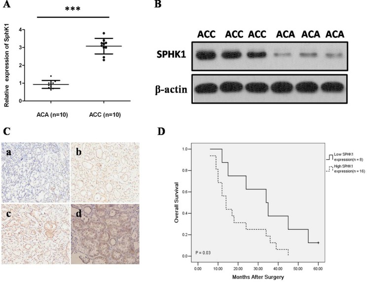
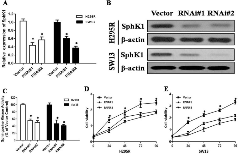
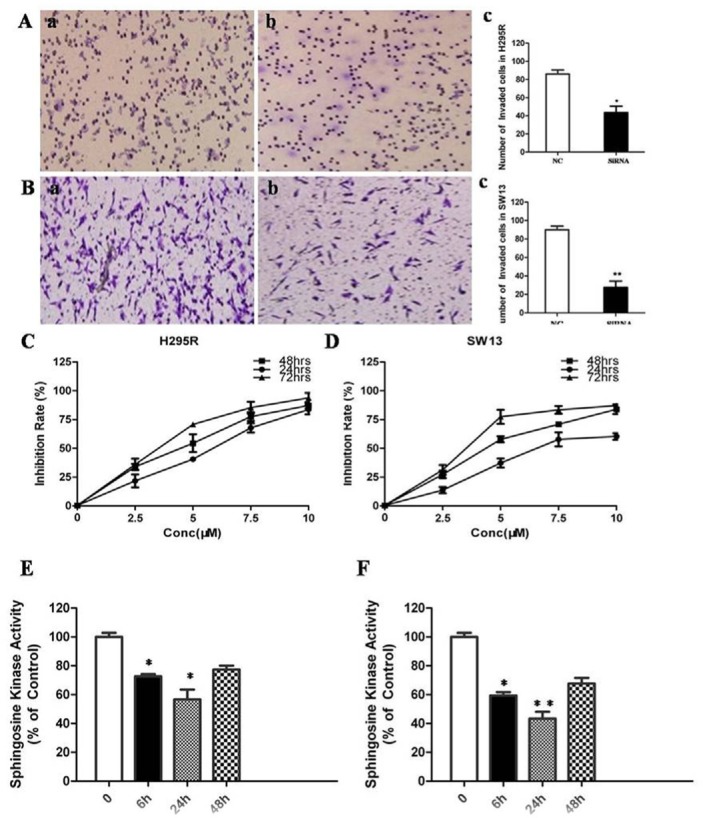
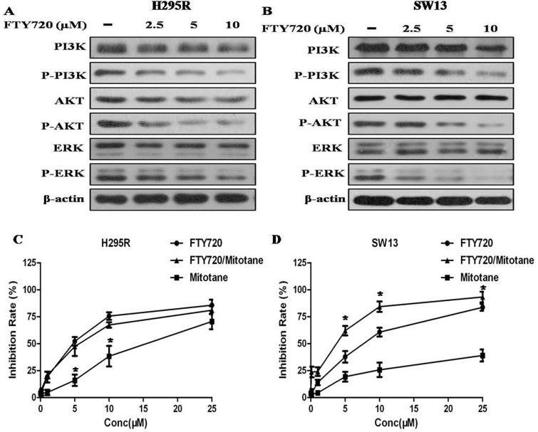
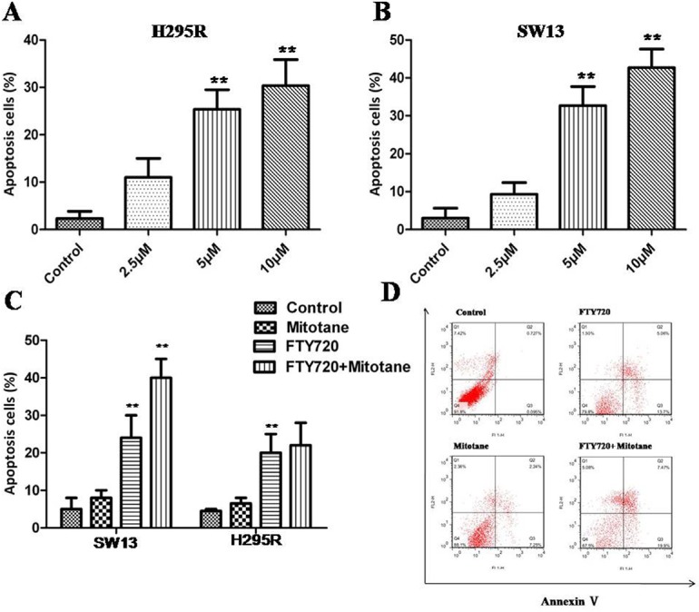
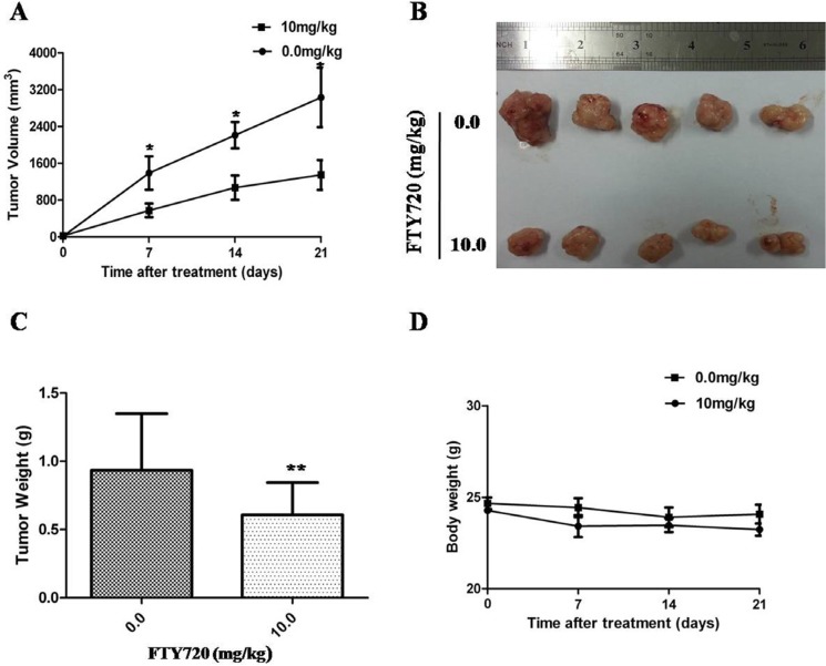
Adrenocortical carcinoma (ACC) is a rare endocrine tumor with a very poor prognosis. Sphingosine kinase 1 (SphK1), an oncogenic kinase, has previously been found to be upregulated in various cancers. However, the role of the SphK1 in ACC has not been investigated. In this study, SphK1 mRNA and protein expression levels as well as clinicopathological significance were evaluated in ACC samples. In vitro siRNA knockdown of SphK1 in two ACC cell lines (H295R and SW13) was used to determine its effect on cellular proliferation and invasion. In addition, we further evaluated the effect of SphK1 antagonist fingolimod (FTY720) in ACC in vitro and in vivo, as a single agent or in combination with mitotane, and attempted to explore its anticarcinogenic mechanisms. Our results show a significant over-expression of SphK1 mRNA and protein expression in the carcinomas compared with adenomas (P < 0.01 for all comparisons). Functionally, knockdown of SphK1 gene expression in ACC cell lines significantly decreased cell proliferation and invasion. FTY720 could result in a decreased cell proliferation and induction of apoptosis, and the combination of mitotane and FTY720 resulted in a greater anti-proliferative effect over single agent treatment in SW13 cells. Furthermore, FTY720 could markedly inhibit tumor growth in ACC xenografts. SphK1 expression is functionally associated to cellular proliferation, apoptosis, invasion and mitotane sensitivity of ACC. Our data suggest that SphK1 might be a potential therapeutic target for the treatment of ACC.
Keywords: FTY720; adrenocortical carcinoma; prognosis; progression; sphingosine kinase 1.
Conflict of interest statement
The authors disclose no potential conflicts of interest.
Figures
Similar articles
-
Lipoprotein-Free Mitotane Exerts High Cytotoxic Activity in Adrenocortical Carcinoma.J Clin Endocrinol Metab. 2015 Aug;100(8):2890-8. doi: 10.1210/JC.2015-2080. Epub 2015 Jun 29. J Clin Endocrinol Metab. 2015. PMID: 26120791
-
Sphingosine kinase 1 is a potential therapeutic target for nasopharyngeal carcinoma.Oncotarget. 2016 Dec 6;7(49):80586-80598. doi: 10.18632/oncotarget.13014. Oncotarget. 2016. PMID: 27811359 Free PMC article.
-
TOP2A is overexpressed and is a therapeutic target for adrenocortical carcinoma.Endocr Relat Cancer. 2013 May 21;20(3):361-70. doi: 10.1530/ERC-12-0403. Print 2013 Jun. Endocr Relat Cancer. 2013. PMID: 23533247 Free PMC article.
-
Xenograft models for preclinical drug testing: implications for adrenocortical cancer.Mol Cell Endocrinol. 2012 Mar 31;351(1):71-7. doi: 10.1016/j.mce.2011.09.043. Epub 2011 Oct 26. Mol Cell Endocrinol. 2012. PMID: 22056412 Review.
-
New perspectives for mitotane treatment of adrenocortical carcinoma.Best Pract Res Clin Endocrinol Metab. 2020 May;34(3):101415. doi: 10.1016/j.beem.2020.101415. Epub 2020 Mar 5. Best Pract Res Clin Endocrinol Metab. 2020. PMID: 32179008 Review.
Cited by
-
The Role of Biomarkers in Adrenocortical Carcinoma: A Review of Current Evidence and Future Perspectives.Biomedicines. 2021 Feb 10;9(2):174. doi: 10.3390/biomedicines9020174. Biomedicines. 2021. PMID: 33578890 Free PMC article. Review.
-
Elimination of HER3-expressing breast cancer cells using aptamer-siRNA chimeras.Exp Ther Med. 2019 Oct;18(4):2401-2412. doi: 10.3892/etm.2019.7753. Epub 2019 Jul 9. Exp Ther Med. 2019. PMID: 31555351 Free PMC article.
-
Apoptosis regulation in adrenocortical carcinoma.Endocr Connect. 2019 May 1;8(5):R91-R104. doi: 10.1530/EC-19-0114. Endocr Connect. 2019. PMID: 30978697 Free PMC article. Review.
-
Sphingosine Kinase Blockade Leads to Increased Natural Killer T Cell Responses to Mantle Cell Lymphoma.Cells. 2020 Apr 21;9(4):1030. doi: 10.3390/cells9041030. Cells. 2020. PMID: 32326225 Free PMC article.
-
Sphingosine-1-phosphate suppresses chondrosarcoma metastasis by upregulation of tissue inhibitor of metalloproteinase 3 through suppressing miR-101 expression.Mol Oncol. 2017 Oct;11(10):1380-1398. doi: 10.1002/1878-0261.12106. Epub 2017 Aug 8. Mol Oncol. 2017. PMID: 28672103 Free PMC article.
References
-
- Kerkhofs TM, Ettaieb MH, Hermsen IG, Haak HR. Developing treatment for adrenocortical carcinoma. Endocr Relat Cancer. 2015;22:R325–338. - PubMed
-
- Mihai R. Diagnosis, treatment and outcome of adrenocortical cancer. Br J Surg. 2015;102:291–306. - PubMed
-
- Fassnacht M, Terzolo M, Allolio B, Baudin E, Haak H, Berruti A, Welin S, Schade-Brittinger C, Lacroix A, Jarzab B, Sorbye H, Torpy DJ, Stepan V, et al. Combination chemotherapy in advanced adrenocortical carcinoma. N Engl J Med. 2012;366:2189–2197. - PubMed
-
- Schiefler C, Piontek G, Doescher J, Schuettler D, Miβlbeck M, Rudelius M, Haug A, Reiter R, Brockhoff G, Pickhard A. Inhibition of SphK1 reduces radiation-induced migration and enhances sensitivity to cetuximab treatment by affecting the EGFR / SphK1 crosstalk. Oncotarget. 2014;5:9877–9888. doi: 10.18632/oncotarget.2436. - DOI - PMC - PubMed
Publication types
MeSH terms
Substances
LinkOut - more resources
Full Text Sources
Other Literature Sources
