

Usefulness of sonography in the diagnosis of rheumatoid hand
- PMID: 26673521
- PMCID: PMC4603217
- DOI: 10.15557/JoU.2013.0033
Usefulness of sonography in the diagnosis of rheumatoid hand
Abstract
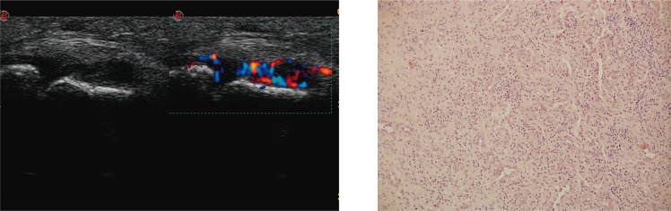
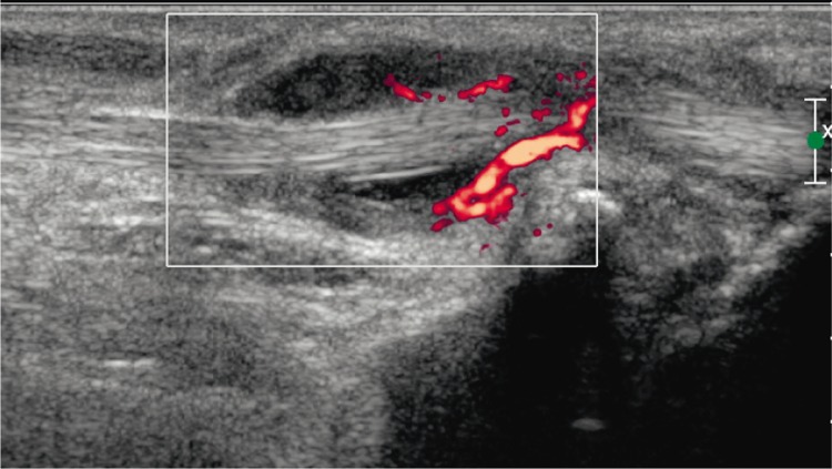
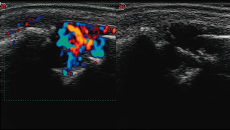
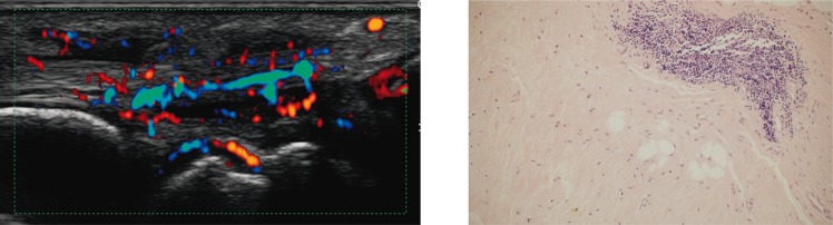
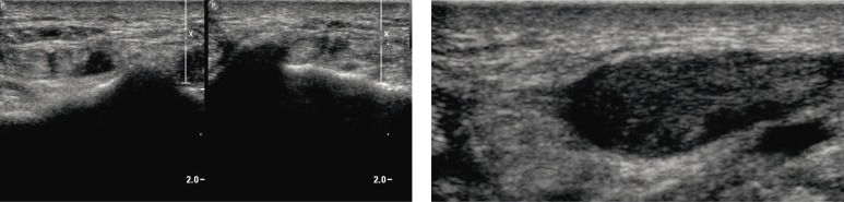
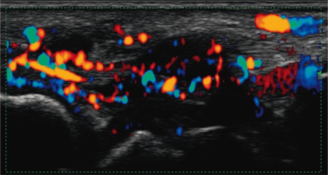
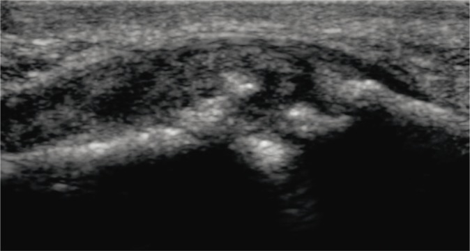
Ultrasound examination is becoming more and more common in patients with rheumatoid diseases. Above all, it enables the assessment of articular soft tissues and constitutes a non-invasive examination. In a rheumatologist's everyday practice, it is conducted at the stage of initial diagnosis as well as to monitor the treatment and to confirm the remission if the clinical picture is ambiguous. The first sign of arthritis (including rheumatoid arthritis) that is visible on ultrasound examination is the thickening of the synovial membrane of the joint cavities, tendon sheaths or bursae. It is frequently accompanied by the exudate in the joint, sheath or bursa. In a subsequent stage, in Doppler examination, enhanced vascularization of the synovial membrane is observed. Sometimes, the inflammatory process of the tendon sheaths also affects the tendons, which might lead to their damage. Moreover, ultrasound examination also reveals erosions and inflammatory cysts (geodes) which attest to the advancement of the disease. A dynamic ultrasound examination enables to diagnose the capsule-ligamentous contracture of the interphalangeal joints, which occurs due to the lack of rehabilitation that should begin at the moment of the commencement of the inflammation. The ultrasound image does not allow for the differentiation between various rheumatoid entities, including those encompassing the joints in the hand, wrist. The observed changes, i.e. thickening of the synovial membrane, hyperemia, effusions, erosions or tendon damage, may accompany various rheumatoid entities. The purpose of the ultrasound examination is to recognize these irregularities, determine their localization and advancement and, finally, to monitor the course of treatment. Furthermore, ultrasound scan enables to assess the joints and tendons in a dynamic examination in relation to local ailments of the patient as well as to monitor the biopsy, aspiration and medicine administration. Sonography is used for a US-guided administration of radioisotope substances for synoviorthesis.
Badanie ultrasonograficzne jest coraz powszechniej wykonywane u pacjentów z chorobami reumatycznymi. Umożliwia ocenę przede wszystkim tkanek miękkich stawów. Ma nieinwazyjny charakter. W codziennej praktyce reumatologa przeprowadza się je na etapie diagnozy, monitorowania leczenia oraz w celu potwierdzenia remisji, jeżeli obraz kliniczny nie jest jednoznaczny. Pierwszym objawem zapalenia stawów (w tym reumatoidalnego zapalenia stawów) widocznym w badaniu ultrasonograficznym jest pogrubienie błony maziowej jam stawów, pochewek lub kaletek. Często towarzyszy mu wysięk w stawie, pochewce lub w kaletce. W kolejnym etapie w badaniu dopplerowskim obserwowane jest wzmożone unaczynienie błony maziowej. Niekiedy proces zapalny pochewek ścięgnistych obejmuje także ścięgna, co może prowadzić do ich uszkodzenia. W badaniu ultrasonograficznym widoczne są ponadto nadżerki i geody, będące dowodem zaawansowania choroby. Dynamiczne badanie ultrasonograficzne pozwala na rozpoznanie przykurczu struktur torebkowo-więzadłowych stawów międzypaliczkowych, najczęściej z powodu braku rehabilitacji, która powinna być rozpoczęta w momencie wystąpienia zapalenia stawu. Obraz ultrasonograficzny nie pozwala na różnicowanie poszczególnych jednostek reumatycznych, w tym obejmujących stawy ręki, także nadgarstka. Obserwowane zmiany, tj. pogrubienie błony maziowej, jej przekrwienie, wysięki, nadżerki, uszkodzenia ścięgien, towarzyszą wielu jednostkom reumatycznym. Celem badania ultrasonograficznego jest rozpoznanie tych nieprawidłowości, określenie ich lokalizacji i zaawansowania, wreszcie monitorowanie w trakcie leczenia. Badanie ultrasonograficzne umożliwia ponadto ocenę stawów i ścięgien w badaniu dynamicznym, w konfrontacji z dolegliwościami punktowymi pacjenta, monitorowanie biopsji, aspiracji, podawania leków. Pod kontrolą ultrasonografii podawane są do stawów radioizotopy w celu synowiortezy radioizotopowej.
Keywords: bursitis; rheumatoid arthritis; rheumatoid hand; sonography; synovitis; tenosynovitis.
Figures
Similar articles
-
Rheumatoid arthritis: sequences.Eur J Radiol. 1998 May;27 Suppl 1:S31-8. doi: 10.1016/s0720-048x(98)00040-0. Eur J Radiol. 1998. PMID: 9652499
-
Ultrasonography for diagnosis, monitoring and treatment of tenosynovitis in patients with rheumatoid arthritis.Dan Med J. 2018 Mar;65(3):B5474. Dan Med J. 2018. PMID: 29510818 Review.
-
The clinical features of rheumatoid arthritis.Eur J Radiol. 1998 May;27 Suppl 1:S18-24. doi: 10.1016/s0720-048x(98)00038-2. Eur J Radiol. 1998. PMID: 9652497 Review.
-
[Technique and diagnostic value of musculoskeletal ultrasonography in rheumatology. Part 6: ultrasonography of the wrist/hand].Z Rheumatol. 2002 Dec;61(6):674-87. doi: 10.1007/s00393-002-0386-6. Z Rheumatol. 2002. PMID: 12491131 German.
-
Ultrasound-detected activity in rheumatoid arthritis on methotrexate therapy: Which joints and tendons should be assessed to predict unstable remission?Rheumatol Int. 2016 Mar;36(3):387-96. doi: 10.1007/s00296-015-3409-8. Epub 2015 Dec 28. Rheumatol Int. 2016. PMID: 26712373
Cited by
-
Ultrasound evaluation of the scapholunate ligament and scapholunate joint space in patients with wrist complaints in a rheumatologic setting.J Ultrason. 2021 Jun 7;21(85):e105-e111. doi: 10.15557/JoU.2021.0019. Epub 2021 Jun 18. J Ultrason. 2021. PMID: 34258035 Free PMC article.
-
High-resolution ultrasound in the evaluation of musculoskeletal infections.J Ultrason. 2023 Nov 23;23(95):e272-e284. doi: 10.15557/jou.2023.0034. eCollection 2023 Oct. J Ultrason. 2023. PMID: 38020512 Free PMC article.
-
Sonography of pathological changes in the hand.J Ultrason. 2014 Mar;14(56):74-88. doi: 10.15557/JoU.2014.0007. Epub 2014 Mar 30. J Ultrason. 2014. PMID: 26675521 Free PMC article. Review.
-
Wrist ultrasound examination - scanning technique and ultrasound anatomy. Part 2: Ventral wrist.J Ultrason. 2017 Jun;17(69):123-128. doi: 10.15557/JoU.2017.0018. Epub 2017 Jun 30. J Ultrason. 2017. PMID: 28856021 Free PMC article. Review.
-
Role of ultrasonography in the diagnosis of rheumatic diseases in light of ACR/EULAR guidelines.J Ultrason. 2016 Mar;16(64):55-64. doi: 10.15557/JoU.2016.0006. Epub 2016 Mar 29. J Ultrason. 2016. PMID: 27104003 Free PMC article. Review.
References
-
- Martinoli C, Bianchi S. Nadgarstek. In: Bianchi S, Martinoli C, editors. Ultrasonografia układu mięśniowo-szkieletowego. Vol. 1. Warszawa: MediPage; 2008. pp. 425–494.
Publication types
LinkOut - more resources
Full Text Sources
Other Literature Sources