

Transforming Growth Factor Beta 1 Modulates the Functional Expression of the Neurokinin-1 Receptor in Human Keratocytes
- PMID: 26673553
- PMCID: PMC4989870
- DOI: 10.3109/02713683.2015.1088954
Transforming Growth Factor Beta 1 Modulates the Functional Expression of the Neurokinin-1 Receptor in Human Keratocytes
Abstract
Purpose: Transforming growth factor beta 1 (TGF-β1) is a cytokine involved in a variety of processes, such as differentiation of fibroblasts into myofibroblasts. TGF-β1 has also been shown to delay the internalization of the neurokinin-1 receptor (NK-1 R) after its activation by its ligand, the neuropeptide substance P (SP). NK-1 R comprises two naturally occurring variants, a full-length and a truncated form, triggering different cellular responses. SP has been shown to affect important events in the cornea - such as stimulating epithelial cell proliferation - processes that are involved in corneal wound healing and thus in maintaining the transparency of the corneal stroma. An impaired signaling through NK-1 R could thus impact the visual quality. We hypothesize that TGF-β1 modulates the expression pattern of NK-1 R in human corneal stroma cells, keratocytes. The purpose of this study was to test that hypothesis.
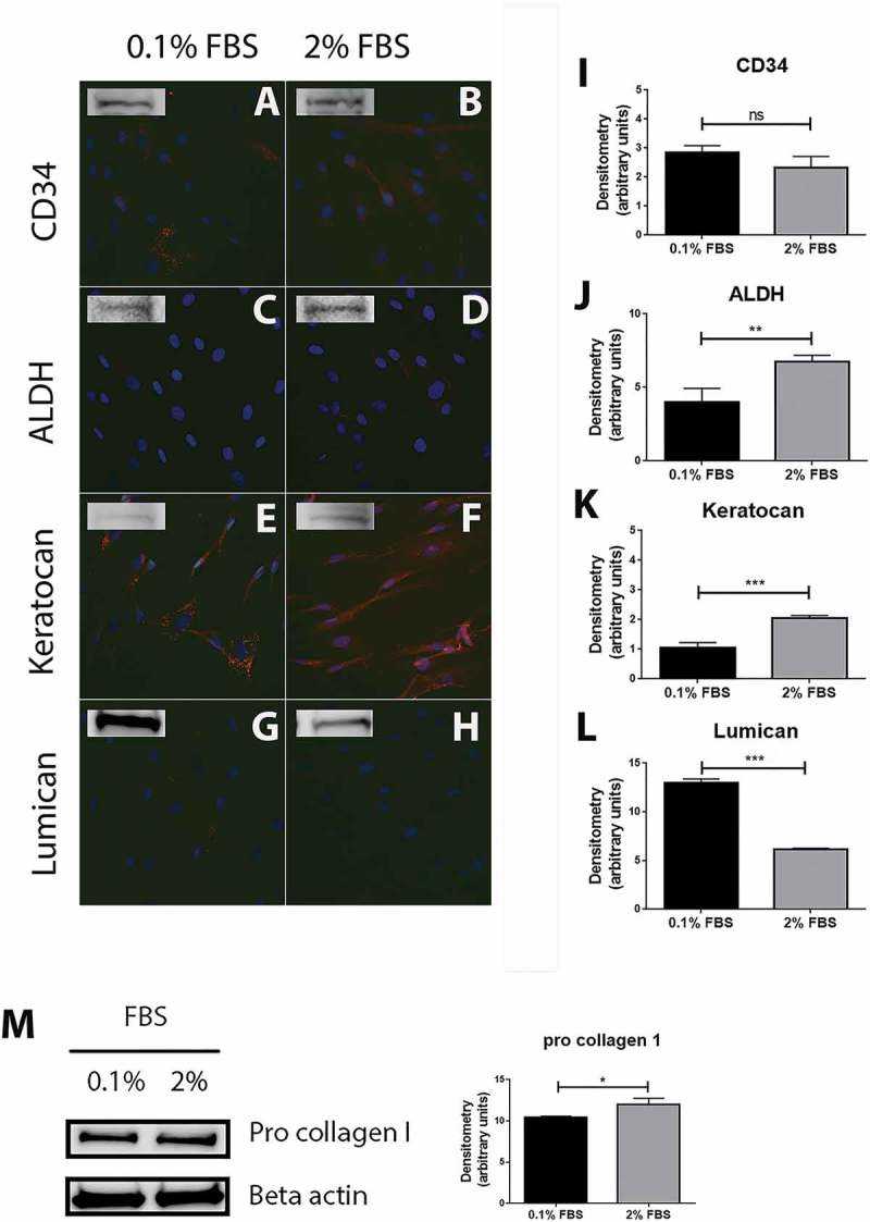
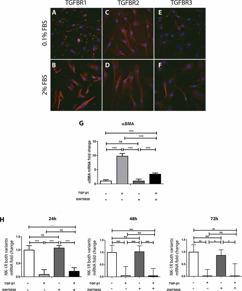
Methods: Cultures of primary keratocytes were set up with cells derived from healthy human corneas, obtained from donated transplantation graft leftovers, and characterized by immunocytochemistry and Western blot. Immunocytochemistry for TGF-β receptors and NK-1 R was performed. Gene expression was assessed with real-time polymerase chain reaction (qPCR).
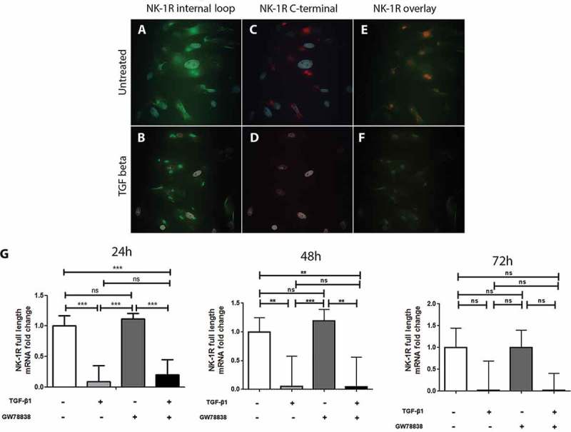
Results: Expression of TGF-β receptors was confirmed in keratocytes in vitro. Treating the cells with TGF-β1 significantly reduced the gene expression of NK-1 R. Furthermore, immunocytochemistry for NK-1 R demonstrated that it is specifically the expression of the full-length isotype of the receptor that is reduced after treatment with TGF-β1, which was also confirmed with qPCR using a specific probe for the full-length receptor.
Conclusions: TGF-β1 down-regulates the gene expression of the full-length variant of NK-1 R in human keratocytes, which might impact its signaling pathway and thus explain the known delay in internalization after activation by SP seen with TGF-β1 treatment.
Keywords: Cornea; cytokines; neuropeptides; stroma; substance P.
Figures
Similar articles
-
IL-1 and TGF-β Modulation of Epithelial Basement Membrane Components Perlecan and Nidogen Production by Corneal Stromal Cells.Invest Ophthalmol Vis Sci. 2018 Nov 1;59(13):5589-5598. doi: 10.1167/iovs.18-25202. Invest Ophthalmol Vis Sci. 2018. PMID: 30480706 Free PMC article.
-
Expression of insulin-like growth factor 2 receptor in corneal keratocytes during differentiation and in response to wound healing.Invest Ophthalmol Vis Sci. 2014 Oct 30;55(12):7697-708. doi: 10.1167/iovs.14-15179. Invest Ophthalmol Vis Sci. 2014. PMID: 25358730 Free PMC article.
-
Downregulation of IL-7 and IL-7R Reduces Membrane-Type Matrix Metalloproteinase 14 in Granular Corneal Dystrophy Type 2 Keratocyte.Invest Ophthalmol Vis Sci. 2018 Nov 1;59(13):5693-5703. doi: 10.1167/iovs.18-25161. Invest Ophthalmol Vis Sci. 2018. PMID: 30489629
-
FGF-2- and TGF-β1-induced downregulation of lumican and keratocan in activated corneal keratocytes by JNK signaling pathway.Invest Ophthalmol Vis Sci. 2011 Nov 21;52(12):8957-64. doi: 10.1167/iovs.11-8078. Invest Ophthalmol Vis Sci. 2011. PMID: 22025571 Free PMC article.
-
The corneal fibrosis response to epithelial-stromal injury.Exp Eye Res. 2016 Jan;142:110-8. doi: 10.1016/j.exer.2014.09.012. Exp Eye Res. 2016. PMID: 26675407 Free PMC article. Review.
Cited by
-
Gatifloxacin inducing apoptosis of stromal fibroblasts through cross-talk between caspase-dependent extrinsic and intrinsic pathways.Int J Ophthalmol. 2019 Oct 18;12(10):1524-1530. doi: 10.18240/ijo.2019.10.02. eCollection 2019. Int J Ophthalmol. 2019. PMID: 31637186 Free PMC article.
-
Seeding of breast cancer cell line (MDA-MB-231LUC+) to the mandible induces overexpression of substance P and CGRP throughout the trigeminal ganglion and widespread peripheral sensory neuropathy throughout all three of its divisions.Mol Pain. 2021 Jan-Dec;17:17448069211024082. doi: 10.1177/17448069211024082. Mol Pain. 2021. PMID: 34229504 Free PMC article.
-
The effects of substance P and acetylcholine on human tenocyte proliferation converge mechanistically via TGF-β1.PLoS One. 2017 Mar 16;12(3):e0174101. doi: 10.1371/journal.pone.0174101. eCollection 2017. PLoS One. 2017. PMID: 28301610 Free PMC article.
-
Cross-talk between the transcription factor Sp1 and C/EBPβ modulates TGFβ1 production to negatively regulate the expression of chemokine RANTES.Heliyon. 2018 Jul 4;4(7):e00679. doi: 10.1016/j.heliyon.2018.e00679. eCollection 2018 Jul. Heliyon. 2018. PMID: 29998198 Free PMC article.
References
-
- Liu C-Y, Birk DE, Hassell JR, Kane B, Kao WW-Y. Keratocan-deficient mice display alterations in corneal structure. J Biol Chem. 2003 Jun 13;278(24):21672–21677. - PubMed
-
- Tuominen IS, Tervo TM, Teppo AM, Valle TU, Grönhagen-Riska C, Vesaluoma MH. Human tear fluid PDGF-BB, TNF-alpha and TGF-beta1 vs corneal haze and regeneration of corneal epithelium and subbasal nerve plexus after PRK. Exp Eye Res. 2001 Jun;72(6):631–641. - PubMed
Publication types
MeSH terms
Substances
LinkOut - more resources
Full Text Sources
Other Literature Sources