

doi: 10.15557/JoU.2014.0048.
Epub 2014 Dec 30.
Why consider contrast-enhanced ultrasound (ce-US) in children?: Editorial comment on: M.M. Woźniak, A. Pawelec, A.P. Wieczorek, M.M. Zajączkowska, H. Borzęcka and P. Nachulewicz 2D/3D/4D contrast-enhanced voiding urosnography in the diagnosis and monitoring of treatment of vesicoureteral reflux in children - can it replace voiding cystourethrography?
Affiliations
- PMID: 26673607
- PMCID: PMC4579718
- DOI: 10.15557/JoU.2014.0048
Item in Clipboard
Why consider contrast-enhanced ultrasound (ce-US) in children?: Editorial comment on: M.M. Woźniak, A. Pawelec, A.P. Wieczorek, M.M. Zajączkowska, H. Borzęcka and P. Nachulewicz 2D/3D/4D contrast-enhanced voiding urosnography in the diagnosis and monitoring of treatment of vesicoureteral reflux in children - can it replace voiding cystourethrography?
J Ultrason.
2014 Dec.
No abstract available
Figures

Ce-VUS for detection of VUR. Serial images demonstrate the contrast reflux into the distal ureter in a oblique section (A) – where a second ureter also seems to be present and refluxing (equalling low pressure VUR of mild degree during filling), one contrast filled mid ureter portion (B), and the dynamic contrast reflux into the renal collecting system (C), with increasing dilatation during voiding (D) indicating additional dilating high grade and high pressure VUR. Also note the duplication of the collecting system in C and D
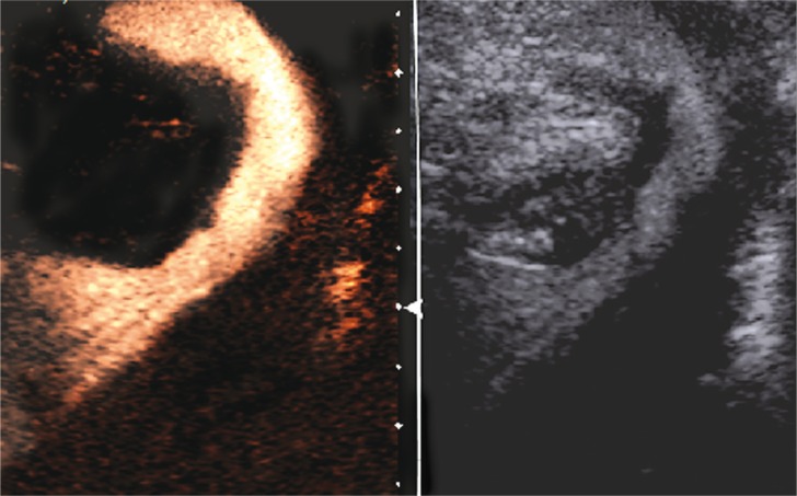
Ce-VUS – urethra assessment during voiding using a perineal approach. At the end of ce-VUS, the bladder is emptied and the urethra is visualised from a perineal approach. Dual image technique: left – contrast dedicated image, right – orienting grey scale b-mode image

Intravenous ce-US (CEUS) of the liver in a treated and already shrunken, partially calicafied liver haemangiendothelioma (A) with no central but little peripheral enhancement in early phases (B, dual image technique), but some residual peripheral slightly nodular enhancement on late scans (C, between + … + ). Additionally for demonstration of CEUS images and effects: normal and homogenuous enhancement of spleen (D) and gas bubbles in the liver (similar to portal pneumatosis) as known to happen with ce-US, when bowel is withing the insonated field (E) after the ce-US investigation

US demonstrates also non-contrasted structures, e.g. non-refluxing megaureter (U) behind the contrast filled urinary bladder in a axial view (A), or the regionally dysplastic renal parenchyma in the respective hydronephrotic kidney without any refluxed contrast in the dilated collecting system – proving absence of VUR (longitudinal section) (B)
Similar articles
-
2D/3D/4D contrast-enhanced voiding urosonography in the diagnosis and monitoring of treatment of vesicoureteral reflux in children - can it replace voiding cystourethrography?J Ultrason. 2013 Dec;13(55):394-407. doi: 10.15557/JoU.2013.0042. Epub 2013 Dec 30. J Ultrason. 2013. PMID: 26674600 Free PMC article.
-
Two-dimensional (2D), three-dimensional static (3D) and real-time (4D) contrast enhanced voiding urosonography (ceVUS) versus voiding cystourethrography (VCUG) in children with vesicoureteral reflux.Eur J Radiol. 2016 Jun;85(6):1238-45. doi: 10.1016/j.ejrad.2015.11.006. Epub 2015 Nov 5. Eur J Radiol. 2016. PMID: 26597418
-
3D/4D contrast-enhanced urosonography (ceVUS) in children - is it superior to the 2D technique?J Ultrason. 2018;18(73):120-125. doi: 10.15557/JoU.2018.0017. J Ultrason. 2018. PMID: 30335920 Free PMC article.
-
Imaging for Vesicoureteral Reflux and Ureteropelvic Junction Obstruction.Eur Urol Focus. 2016 Jun;2(2):130-138. doi: 10.1016/j.euf.2016.03.015. Epub 2016 Apr 13. Eur Urol Focus. 2016. PMID: 28723527 Review.
-
Pediatric voiding cystourethrography: An essential examination for urologists but a terrible experience for children.Int J Urol. 2019 Feb;26(2):160-171. doi: 10.1111/iju.13881. Epub 2018 Dec 19. Int J Urol. 2019. PMID: 30569659 Review.
References
-
- Brenner DJ, Hall EJ. Computed tomography – an increasing source of radiation exposure. N Engl J Med. 2007;357:2277–2284. - PubMed
-
- Brenner D, Elliston C, Hall E, Berdon W. Estimated risks of radiationinduced fatal cancer from pediatric CT. AJR Am J Roentgenol. 2001;176:289–296. - PubMed
-
- Committee on the Effects of Atomic Radiation UNSCEAR. Report to the General Assembly with Scientific Annexes: Sources and Effects of Ionizing Radiation; 2008. Available from: http://www.unscear.org/docs/reports/2008/09-86753_Report_2008_Annex_A.pdf.
-
- Image Gently – the Alliance for Radiation Safety in Pediatric Imaging; Available from: http://www.pedrad.org/associations/5364/ig.
LinkOut - more resources
Full Text Sources
Other Literature Sources