

Mesenteric changes in an ultrasound examination can facilitate the diagnosis of neuroendocrine tumors of the small intestine
- PMID: 26673610
- PMCID: PMC4657393
- DOI: 10.15557/JoU.2015.0024
Mesenteric changes in an ultrasound examination can facilitate the diagnosis of neuroendocrine tumors of the small intestine
Abstract
Neuroendocrine tumors make up an interesting pathology of a variable clinical picture, prognosis, localization, endocrine activity and degree of malignancy.
Aim: The aim of this paper is to assess whether ultrasonography can be helpful in diagnosing neuroendocrine tumors in the small intestine by analyzing changes in the mesentery.
Material and methods: From 1996 to 2013, we encountered 17 patients (9 women and 8 men at the mean age of 57) with a neuroendocrine tumor in the small intestine. The diagnosis was confirmed in all patients by pathomorphological examinations. All retrospectively analyzed patients (n =17) had an abdominal US examination conducted in accordance with the previously mentioned protocol.
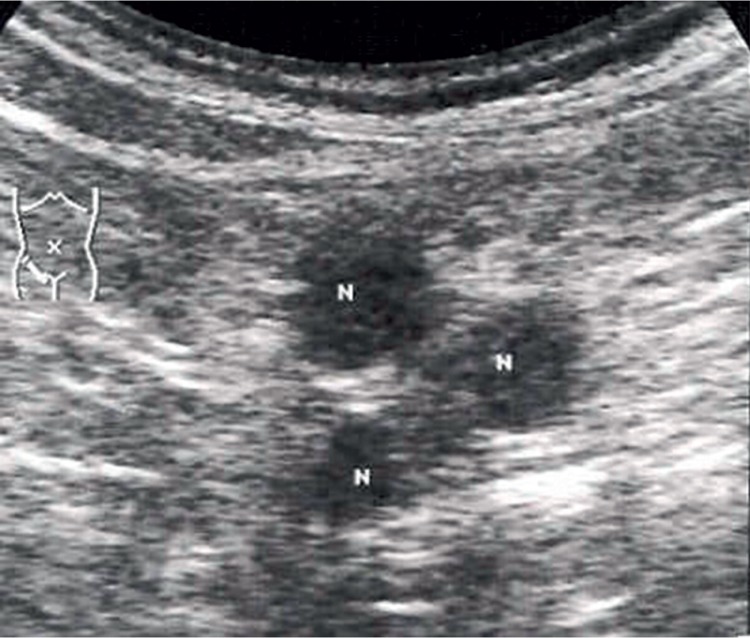
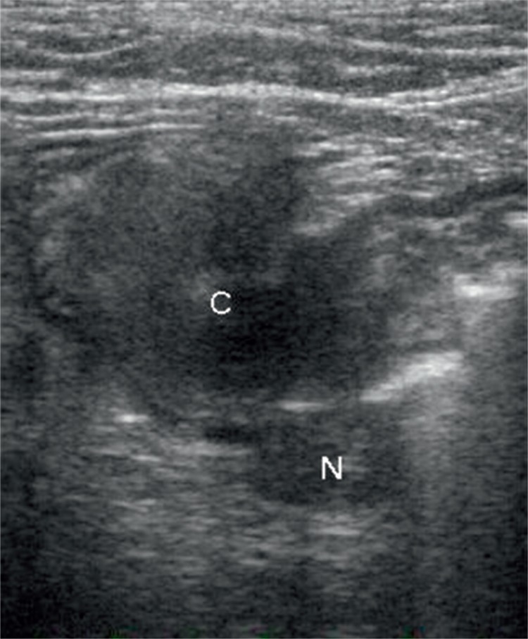
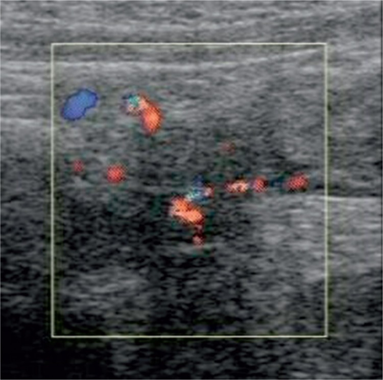
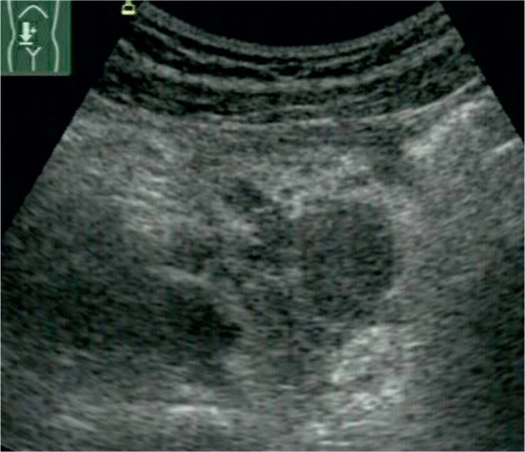
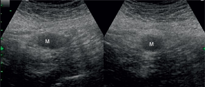
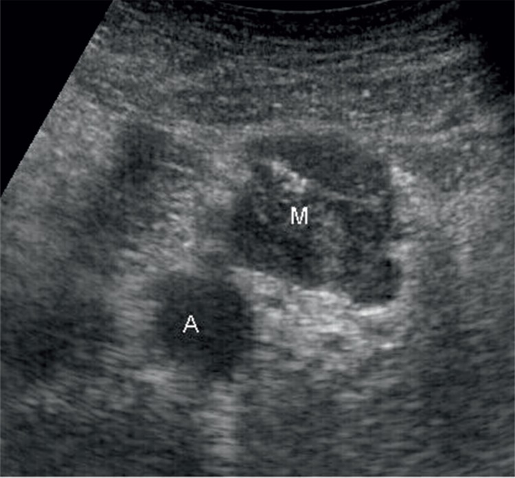
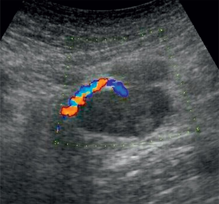
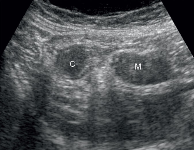
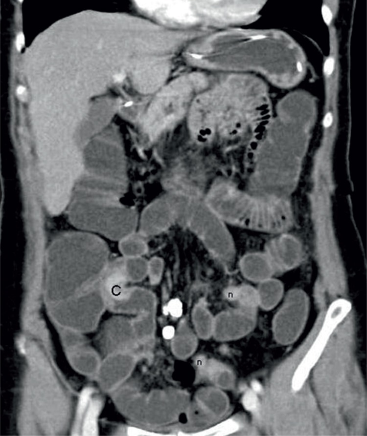
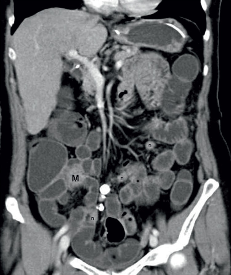
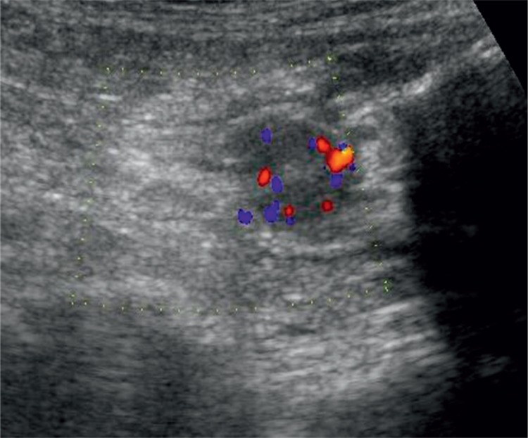
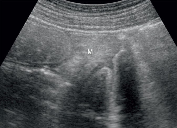
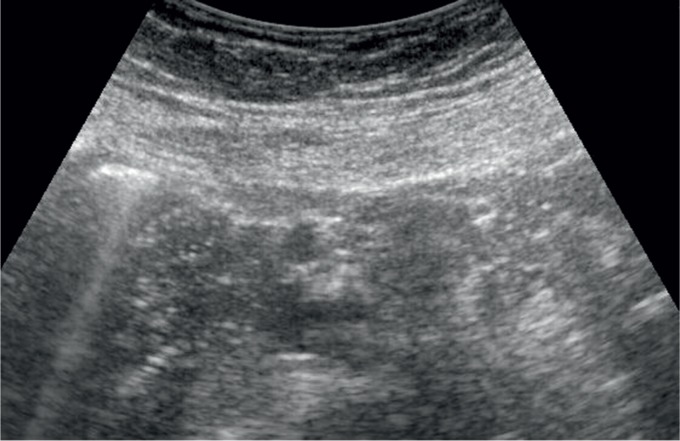
Results: Primary neuroendocrine tumors were visualized ultrasonography in 13 of 17 patients (76.5%). In the remaining 4 patients, tumors in the small bowel were diagnosed in CT enteroclysis (n = 3) and conventional enteroclysis (n = 1). Somatostatin receptor scintigraphy and CT enteroclysis supplemented the ultrasound result by providing information about the stage of the disease in 7 cases. In most of the 17 patients with a neuroendocrine tumor diagnosed by ultrasound (n = 11, 64.7%), changes in the mesentery were prevailing pathological signs that raised suspicion and, consequently, helped localize the primary lesion. The hypoechoic mesenteric lymph nodes were the greatest (9-18 mm), well-circumscribed, and the largest of them showed signs of vascularization. The size of hypoechoic lesions in the mesentery ranged from 25 to 53 mm. These lesions showed moderate blood flow. All of them were single, usually poorly circumscribed; two lesions showed slight calcifications.
Conclusions: In an abdominal US examination, 2/3 of patients with neuroendocrine tumors in the small bowel manifest secondary lesions in the mesentery which are easier to visualize than the primary focus. 30% of them are manifested as hypoechoic masses. The detection of such lesions should prompt the search for the primary focus in the small intestine.
Nowotwory neuroendokrynne to ciekawy dział patologii o zróżnicowanym obrazie klinicznym, rokowaniu, umiejscowieniu, czynności wydzielniczej i stopniu złośliwości.
Cel pracy: Celem pracy jest ocena możliwości diagnostyki nowotworów neuroendokrynnych jelita cienkiego przy użyciu ultrasonografii poprzez identyfikację zmian w krezce.
Materiał i metoda: W okresie od 1996 do 2013 roku zgromadzono 17 chorych (9 kobiet i 8 mężczyzn w średnim wieku 57 lat) z nowotworem neuroendokrynnym w jelicie cienkim. U wszystkich pacjentów potwierdzono rozpoznanie nowotworu neuroendokrynnego na podstawie badań patomorfologicznych. Wszystkie objęte retrospektywną analizą osoby (n = 17) miały wykonane badanie USG jamy brzusznej według protokołu opisanego wcześniej.
Wyniki: W badaniu ultrasonograficznym udało się uwidocznić pierwotny nowotwór neuroendokrynny u 13 z 17 chorych (76,5%). U pozostałych 4 osób guz zlokalizowany w jelicie cienkim został rozpoznany na podstawie enteroklizy TK (n = 3) i enteroklizy klasycznej (n = 1). Somatostatynowa scyntygrafia receptorowa i enterokliza TK dostarczyły dodatkowych informacji co do stopnia zaawansowania procesu chorobowego w stosunku do wyników badania USG w 7 przypadkach. Wśród 17 chorych z rozpoznanym ultrasonograficznie nowotworem neuroendokrynnym jelita cienkiego u większości (n = 11, 64,7%) zmiany w krezce jelita były dominującym objawem patologicznym, który pozwolił podejrzewać, a następnie zlokalizować zmianę pierwotną. Hipoechogeniczne węzły chłonne krezkowe miały największe wymiary (9–18 mm), były dobrze odgraniczone, a większe z nich wykazywały unaczynienie. Wielkość hipoechogenicznej masy w krezce wahała się od 25 do 53 mm. Zmiany tego typu wykazywały umiarkowane unaczynienie. Wszystkie były pojedyncze, przeważnie nieostro odgraniczone, a w dwóch zmianach obecne były niewielkie zwapnienia.
Wnioski: W badaniu USG przezbrzusznym u 2/3 chorych z guzem neuroendokrynnym w jelicie cienkim występują wtórne zmiany w krezce, które są łatwiejsze do zobrazowania niż samo ognisko pierwotne. W 30% manifestują się jako masy hipoechogeniczne. Wykrycie takich zmian powinno skłonić do poszukiwania ogniska pierwotnego w jelicie cienkim.
Keywords: abdominal ultrasound; diagnosis; mesenteric lesions; neuroendocrine tumors; small intestine.
Figures

Similar articles
-
Neuroendocrine tumor of the small intestine diagnosed with trans-abdominal ultrasonography: A case report.Int J Surg Case Rep. 2017;31:75-78. doi: 10.1016/j.ijscr.2017.01.012. Epub 2017 Jan 10. Int J Surg Case Rep. 2017. PMID: 28122317 Free PMC article.
-
Ultrasound of selected pathologies of the small intestine.J Ultrason. 2013 Jun;13(53):155-66. doi: 10.15557/JoU.2013.0016. Epub 2013 Jun 30. J Ultrason. 2013. PMID: 26672622 Free PMC article.
-
Ultrasound in Crohn's disease of the small bowel.Eur J Radiol. 2000 Sep;35(3):176-82. doi: 10.1016/s0720-048x(00)00240-0. Eur J Radiol. 2000. PMID: 11000560
-
Mesenteric neoplasms: CT appearances of primary and secondary tumors and differential diagnosis.Radiographics. 2003 Mar-Apr;23(2):457-73; quiz 535-6. doi: 10.1148/rg.232025081. Radiographics. 2003. PMID: 12640160 Review.
-
[Imaging methods in diagnosis of neuroendocrine tumors of the gastrointestinal tract].Bildgebung. 1995 Mar;62(1):5-13. Bildgebung. 1995. PMID: 7756824 Review. German.
Cited by
-
Management of Gastrointestinal Neuroendocrine Tumors.Clin Med Insights Endocrinol Diabetes. 2019 Oct 24;12:1179551419884058. doi: 10.1177/1179551419884058. eCollection 2019. Clin Med Insights Endocrinol Diabetes. 2019. PMID: 31695546 Free PMC article. Review.
-
Neuroendocrine tumor of the small intestine diagnosed with trans-abdominal ultrasonography: A case report.Int J Surg Case Rep. 2017;31:75-78. doi: 10.1016/j.ijscr.2017.01.012. Epub 2017 Jan 10. Int J Surg Case Rep. 2017. PMID: 28122317 Free PMC article.
References
-
- Kos-Kudła B. Guzy neuroendokrynne przewodu pokarmowego; Gdańsk: Via Medica; 2010.
-
- Modlin IM, Lye KD, Kidd M. A 5-decade analysis of 13,715 carcinoid tumors. Cancer. 2003;97:934–959. - PubMed
-
- Modlin IM, Champoneria MC, Chan AK, Kidd M. A three-decade analysis 3,911 small intestinal neuroendocrine tumors: the rapid pace of no progress. Am J Gastroenterol. 2007;102:1464–1473. - PubMed
-
- Yao JC, Hassan M, Phan A, Dagohoy C, Learg C, Mares JE, et al. One hundred years after “carcinoid”: epidemiology of and prognostic factors for neuroendocrine tumors in 35,825 cases in the United States. J Clin Oncol. 2008;26:3063–3072. - PubMed
LinkOut - more resources
Full Text Sources