. 2015 Dec 17;6(12):e2018.
doi: 10.1038/cddis.2015.360.

Nucleoprotein of influenza A virus negatively impacts antiapoptotic protein API5 to enhance E2F1-dependent apoptosis and virus replication

Affiliations

Nucleoprotein of influenza A virus negatively impacts antiapoptotic protein API5 to enhance E2F1-dependent apoptosis and virus replication

A K Mayank et al. Cell Death Dis. .

Abstract

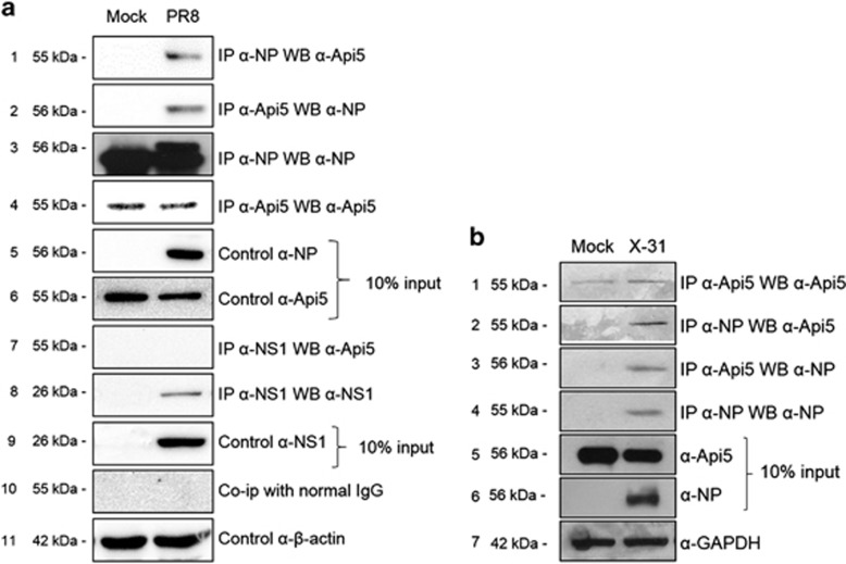
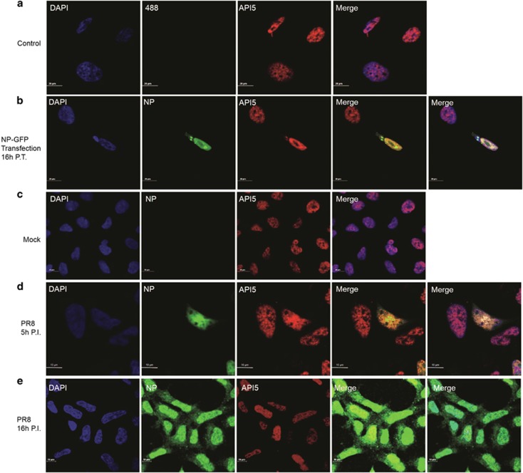
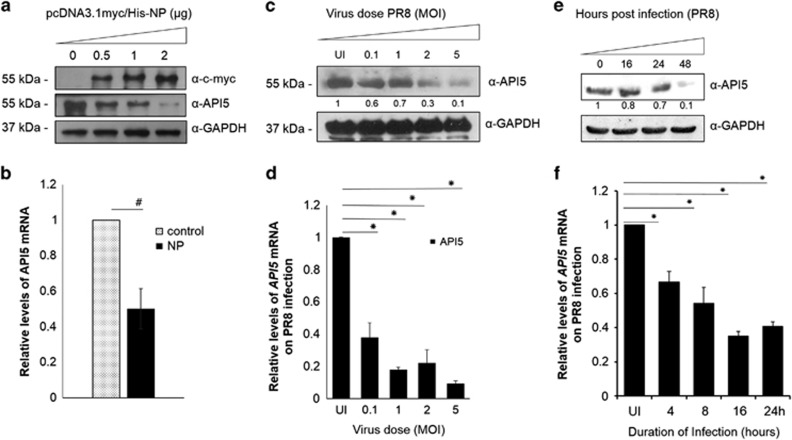
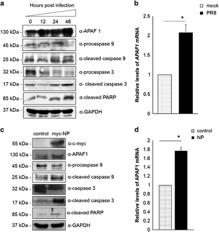
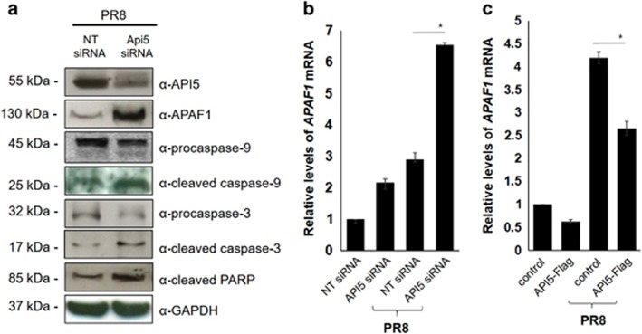
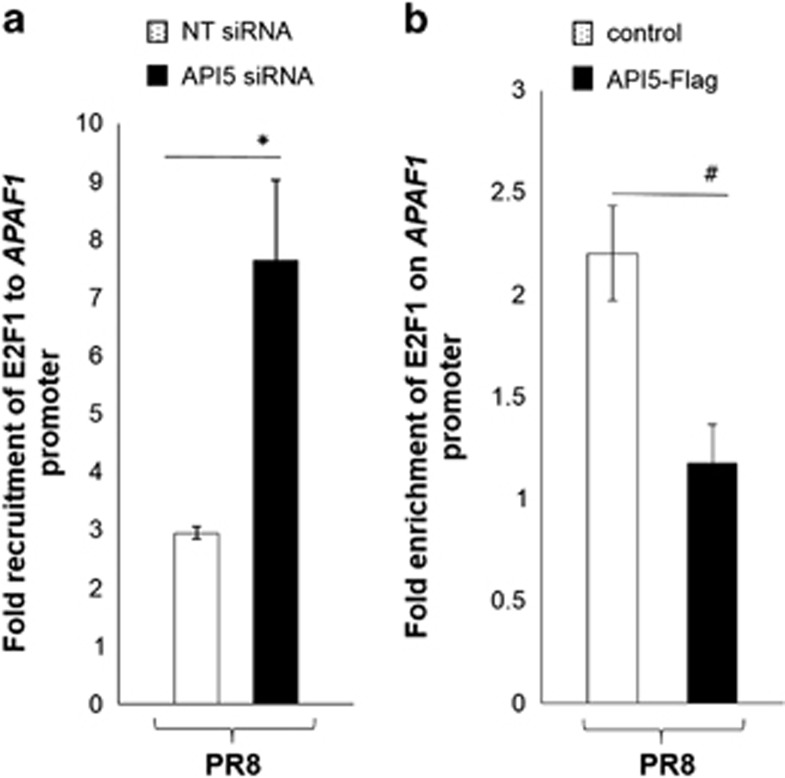
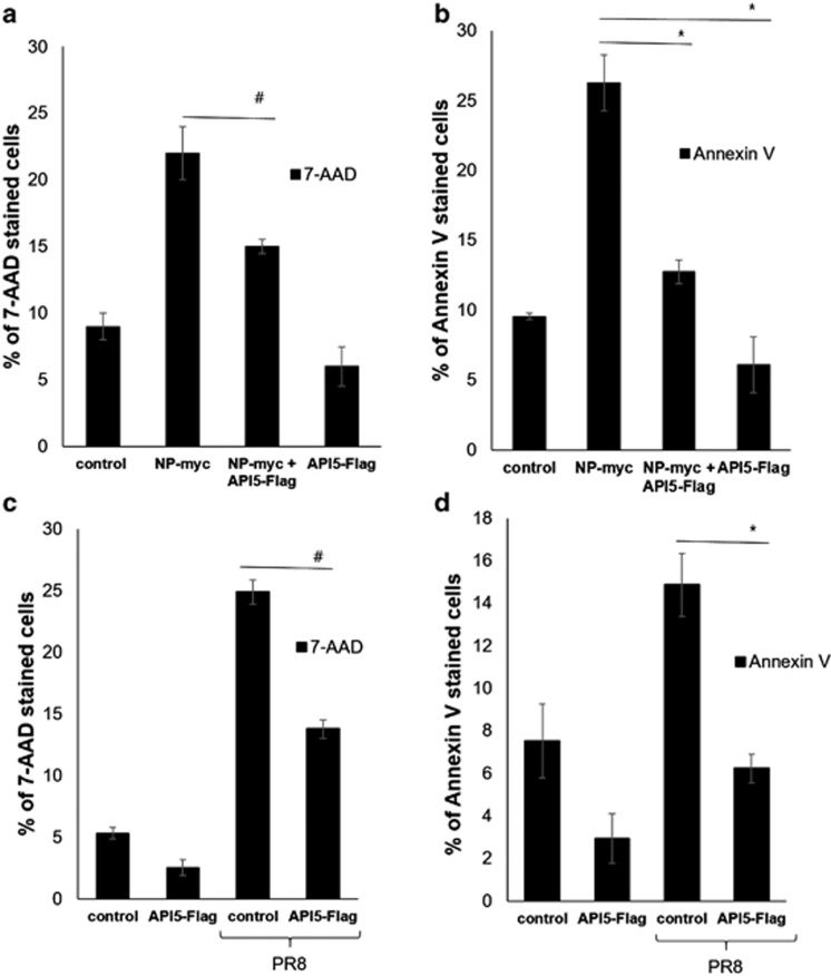
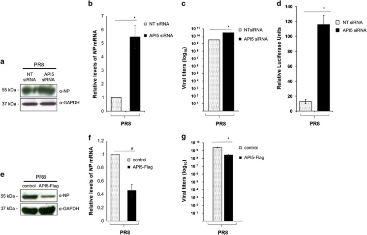
Apoptosis of host cells profoundly influences virus propagation and dissemination, events that are integral to influenza A virus (IAV) pathogenesis. The trigger for activation of apoptosis is regulated by an intricate interplay between cellular and viral proteins, with a strong bearing on IAV replication. Though the knowledge of viral proteins and mechanisms employed by IAV to induce apoptosis has advanced considerably of late, we know relatively little about the repertoire of host factors targeted by viral proteins. Thus, identification of cellular proteins that are hijacked by the virus will help us not only to understand the molecular underpinnings of IAV-induced apoptosis, but also to design future antiviral therapies. Here we show that the nucleoprotein (NP) of IAV directly interacts with and suppresses the expression of API5, a host antiapoptotic protein that antagonizes E2F1-dependent apoptosis. siRNA-mediated depletion of API5, in NP-overexpressed as well as IAV-infected cells, leads to upregulation of apoptotic protease activating factor 1 (APAF1), a downstream modulator of E2F1-mediated apoptosis, and cleavage of caspases 9 and 3, although a reciprocal pattern of these events was observed on ectopic overexpression of API5. In concordance with these observations, annexin V and 7AAD staining assays exhibit downregulation of early and late apoptosis in IAV-infected or NP-transfected cells on overexpression of API5. Most significantly, while overexpression of API5 decreases viral titers, cellular NP protein as well as mRNA levels in IAV-infected A549 cells, silencing of API5 expression causes a steep rise in the same parameters. From the data reported in this manuscript, we propose a proapoptotic role for NP in IAV pathogenesis, whereby it suppresses expression of antiapoptotic factor API5, thus potentiating the E2F1-dependent apoptotic pathway and ensuring viral replication.


Figures

Figure 1
Figure 1
Influenza A virus nucleoprotein interacts with API5 in IAV-infected A549 cells. (a) Lung epithelial A549 cells were infected with PR8 at an MOI of 1. At 16 h post infection, cells were collected and lysed for co-immunoprecipitation assays. Panels 1 and 2 show immunoprecipitation of NP by α-NP followed by western blotting with α-API5 antibody and vice versa. Panels 3 and 4 show immunoprecipitation and western blotting with α-NP and α-API5 antibodies, respectively. Panels 5, 6, 9, and 11 show western blotting with α-NP, α-API5, α-NS1, and α-β-actin antibodies. Panels 7 and 8 show immunoprecipitation with α-NS1 (another IAV protein) followed by western blotting with α-API5 and α-NS1 antibody, respectively. Panel 10 shows the isotype control. (b) Lung epithelial A549 cells were infected with X31 at an MOI of 1. At 24 h post infection, cells were collected and lysed for co-immunoprecipitation assays. Panels 2 and 3 show immunoprecipitation of NP by α-NP followed by western blotting with α-API5 antibody and vice versa. Panels 1 and 4 show immunoprecipitation and western blotting with α-API5 and α-NP antibodies, respectively. Panels 5, 6, and 7 show western blotting with α-NP, α-API5, and α-GAPDH antibodies
Figure 2
Figure 2
NP of IAV colocalizes with cellular API5. (a and b) A549 cells were transfected with either control (pcDNA3.1-myc/His) or NP-GFP (pEGFP-NP) plasmids. Cells were fixed with 2% paraformaldehyde 16 h post transfection (P.T.), followed by immunostaining using rabbit α-API5 primary antibody and Alexa594-conjugated anti-rabbit secondary antibody (red). NP is shown in green and nuclei are stained blue with DAPI. A549 cells were infected with either mock or PR8 at an MOI of 1 and fixed with 2% paraformaldehyde 5 h post infection (P.I.) (d) and 16 h post infection (e). API5 was stained using rabbit α-API5 as primary antibody and anti-rabbit conjugated with Alexa594 as secondary antibody (red). NP was stained with mouse α-NP as primary antibody and anti-mouse conjugated with Alexa488 as secondary antibody (green). Nuclei are stained blue with DAPI. (a–c) have a scale of 20 μm and (d and e) have a scale of 10 μm
Figure 3
Figure 3
NP of IAV suppresses API5 expression. (a) A549 cells were transfected with either control (pcDNA3.1-myc/His) or with increasing concentration of myc-NP (pcDNA3.1-myc/His-NP), 48 h post transfection the whole-cell lysates were resolved on SDS-polyacrylamide gel electrophoresis (SDS-PAGE) for detection of NP, API5, and GAPDH using α-c-myc, α-API5, and α-GAPDH antibodies, respectively. (b) A549 cells were transfected with either control (pcDNA3.1-myc/His) or myc-NP (pcDNA3.1-myc/His-NP), 24 h post transfection the total RNA was isolated for the estimation of API5 mRNA levels using quantitative PCR with specific primers. (c) A549 cells were either mock infected (MI) or infected with PR8 at an MOI of 0.1, 1, 2, and 5 for 24 h. The whole-cell lysates from the samples were resolved on SDS-PAGE for detection of NP, API5, and GAPDH. (d) A549 cells were either MI or infected with PR8 at an MOI of 0.1, 1, 2, and 5 for 24 h. Total RNA was isolated for the estimation of API5 mRNA levels using quantitative PCR with specific primers. (e) A549 cells were infected with PR8 at an MOI of 1 and samples were collected at 0, 16, 24, and 48 h post infection (p.i.). The whole-cell lysates from the samples were resolved on SDS-PAGE for detection of API5 and GAPDH. (f) A549 cells were either MI or infected with PR8 at an MOI of 1 and samples were collected at 4, 8, 16, and 24 p.i. Total RNA was isolated for the estimation of API5 mRNA levels by quantitative PCR using specific primers. Fold change in the expression levels of API5 protein are shown below each panel. The data in b, d, and f are shown as mean±S.D. of three independent experiments. # and * indicate statistically significant difference at P<0.05 and P<0.01, respectively
Figure 4
Figure 4
NP of IAV induces APAF1-dependent cleavage of initiator and effector caspases. (a) A549 cells were infected with PR8 at an MOI of 1. The whole-cell lysates were prepared at 0, 12, 24, and 48 h post infection, and were subjected to western blot analysis using indicated primary antibodies. (b) A549 cells were either mock infected (mock) or infected with PR8 (PR8) at an MOI of 1. At 24 h post infection, the total RNA was isolated for the estimation of APAF1 mRNA levels by quantitative PCR using specific primers. (c) A549 cells were transfected with either control (pcDNA3.1-myc/His) or myc-NP (pcDNA3.1-myc/His-NP), and the whole-cell lysates were prepared at 48 h post transfection. The lysates were subjected to western blot analysis using indicated primary antibodies. (d) A549 cells were transfected with either control (pcDNA3.1-myc/His) or myc-NP (pcDNA3.1-myc/His-NP). At 48 h post transfection, the total RNA was isolated for the estimation of APAF1 mRNA levels by quantitative PCR using specific primers. The data in b and d are shown as mean±S.D. of three independent experiments. *indicates statistically significant difference at P<0.01
Figure 5
Figure 5
API5 promotes antiapoptotic effect in infected cells. A549 cells were transfected with either non-targeting (NT siRNA) or API5-specific siRNA (API5 siRNA). At 24 h post transfection, cells were infected with PR8 at an MOI of 1. At 24 h post infection, (a) the whole-cell lysate was resolved on SDS-polyacrylamide gel electrophoresis for detection of API5, APAF1, PARP, caspases 9 and 3 using indicated primary antibodies. (b) A549 cells were transfected with either NT siRNA or API5 siRNA. At 24 h post transfection, cells were infected with either mock or PR8 at an MOI of 1. At 24 h post infection, the total RNA was isolated for the estimation of APAF1 mRNA by quantitative PCR using specific primers. (c) A549 cells were transfected with either control (pLPC) or API5-Flag (pLPC-API5-Flag), 24 h post transfection cells were infected with either mock or PR8 at an MOI of 1. At 24 h post infection, the total RNA was isolated for the estimation of APAF1 mRNA by quantitative PCR using specific primers. The data in b and c are shown as mean±S.D. of three independent experiments. * indicates statistically significant difference at P<0.01
Figure 6
Figure 6
API5 alters E2F1 recruitment on APAF1 promoter. A549 cells were transfected with (a) either non-targeting siRNA (NT siRNA) or API5-specific siRNA (API5 siRNA), (b) either control (pLPC) or API5-Flag (pLPC-API5-Flag). At 24 h post transfection, cells were infected with PR8 at an MOI of 1. Cells were fixed 24 h post infection for isolation of protein–DNA complexes and immunoprecipitated with E2F1 antibody. The genomic DNA bound to E2F1 antibody was isolated and analyzed by quantitative PCR using APAF1 primers. The data are shown as mean±S.D. of three independent experiments. # and * indicate statistically significant difference at P<0.05 and P<0.01, respectively
Figure 7
Figure 7
API5 suppresses NP-induced apoptosis. (a and b) A549 cells were transfected with either control (pcDNA3.1-myc/His), NP-myc (pcDNA3.1-myc/His-NP), API5-Flag (pLPC-API5-Flag) or co-transfected with NP-myc and API5-Flag. At 48 h post transfection, cells were collected and stained with either FITC-conjugated annexin V or 7AAD and subjected to flow cytometry, and were later analyzed using BD Cell Quest pro software (NJ, USA) and plotted as graphs. (c and d) A549 cells were transfected with either control (pcDNA3.1-myc/His) or API5-Flag (pLPC-API5-Flag). At 24 h post transfection, cells were infected with PR8 at an MOI of 1. Cells were collected 24 h post infection and stained with FITC-conjugated annexin V or 7AAD and subjected to flow cytometry, and were later analyzed using BD Cell Quest and plotted as graphs. For acquisition 20 000 cells per sample were used. The data are shown as mean±S.D. of three independent experiments. # and * indicate statistically significant difference at P<0.05 and P<0.01, respectively
Figure 8
Figure 8
Effect of cellular status of API5 on viral replication. A549 cells were transfected with either non-targeting siRNA (NT siRNA) or API5-specific siRNA (API5 siRNA) for 24 h followed by infection with PR8 virus at an MOI 1. The cells were collected at 24 h post infection. (a) The whole-cell lysate was resolved on SDS-polyacrylamide gel electrophoresis (SDS-PAGE) and western blotted with α-NP and α-GAPDH antibodies, respectively. (b) Total RNA was isolated for the estimation of NP mRNA levels using quantitative PCR with specific primers. (c) A549 cells were transfected with either NT siRNA or API5 siRNA for 24 h followed by infection with X31 virus at an MOI 1. The cells were collected at 24 h post infection. Aliquot of supernatant obtained from IAV-infected cells was used to infect MDCK cells followed by the determination of viral titers using a plaque assay. (d) Plasmids encoding polymerase complex components (PA, PB1, PB2, and NP) derived from PR8 (H1N1 virus) were co-transfected alongside a reporter plasmid containing noncoding sequence from the NS1 segment of influenza A virus and luciferase gene driven by the pol I promoter in A549 cells that had been pretreated with either NT or API5 siRNA. Plasmid pRL-TK (Promega, WI, USA), which expresses renilla luciferase, was co-transfected as an internal control for data normalization. (e) A549 cells were transfected with either control (pcDNA3.1-myc/His) or API5-Flag (pLPC-API5-Flag) for 24 h followed by infection with either mock (allantoic fluid) or PR8 at an MOI 1. The cells were collected at 24 h post infection. The whole-cell lysate was resolved on SDS-PAGE and western blotted with anti-NP and anti-GAPDH antibodies, respectively. (f) Total RNA was isolated for the estimation of NP mRNA levels using quantitative PCR with specific primers. (g) A549 cells were transfected with either control (pcDNA3.1-myc/His) or API5-Flag (pLPC-API5-Flag) for 24 h followed by infection with X31 virus at an MOI 1. The cells were collected at 24 h post infection. Aliquot of supernatant obtained from IAV-infected cells was used to infect MDCK cells followed by the determination of viral titers using a plaque assay. The data in b, c, d, f and g are shown as mean±S.D. of three independent experiments. # and * indicate statistically significant difference at P<0.05 and P<0.01, respectively

Similar articles

Cited by

References

    1. 1O'Brien V. Viruses and apoptosis. J Gen Virol 1998; 79: 1833–1845. - PubMed
    1. 2Benedict CA, Norris PS, Ware CF. To kill or be killed: viral evasion of apoptosis. Nat Immunol 2002; 3: 1013–1018. - PubMed
    1. 3Hardwick JM. Viral interference with apoptosis. Semin Cell Dev Biol 1998; 9: 339–349. - PubMed
    1. 4Bantel H, Schulze-Osthoff K. Apoptosis in hepatitis C virus infection. Cell Death Differ 2003; 10(Suppl 1): S48–S58. - PubMed
    1. 5Tollefson AE, Ryerse JS, Scaria A, Hermiston TW, Wold WS. The E3-11.6-kDa adenovirus death protein (ADP) is required for efficient cell death: characterization of cells infected with adp mutants. Virology 1996; 220: 152–162. - PubMed

Publication types

MeSH terms