Histochemical Detection of Collagen Fibers by Sirius Red/Fast Green Is More Sensitive than van Gieson or Sirius Red Alone in Normal and Inflamed Rat Colon
- PMID: 26673752
- PMCID: PMC4682672
- DOI: 10.1371/journal.pone.0144630
Histochemical Detection of Collagen Fibers by Sirius Red/Fast Green Is More Sensitive than van Gieson or Sirius Red Alone in Normal and Inflamed Rat Colon
Abstract
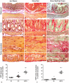
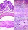
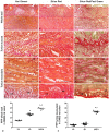
Collagen detection in histological sections and its quantitative estimation by computer-aided image analysis represent important procedures to assess tissue localization and distribution of connective fibers. Different histochemical approaches have been proposed to detect and quantify collagen deposition in paraffin slices with different degrees of satisfaction. The present study was performed to compare the qualitative and quantitative efficiency of three histochemical methods available for collagen staining in paraffin sections of colon. van Gieson, Sirius Red and Sirius Red/Fast Green stainings were carried out for collagen detection and quantitative estimation by morphometric image analysis in colonic specimens from normal rats or animals with 2,4-dinitrobenzenesulfonic acid (DNBS) induced colitis. Haematoxylin/eosin staining was carried out to assess tissue morphology and histopathological lesions. Among the three investigated methods, Sirius Red/Fast Green staining allowed to best highlight well-defined red-stained collagen fibers and to obtain the highest quantitative results by morphometric image analysis in both normal and inflamed colon. Collagen fibers, which stood out against the green-stained non-collagen components, could be clearly appreciated, even in their thinner networks, within all layers of normal or inflamed colonic wall. The present study provides evidence that, as compared with Sirius Red alone or van Gieson staining, the Sirius Red/Fast Green method is the most sensitive, in terms of both qualitative and quantitative evaluation of collagen fibers, in paraffin sections of both normal and inflamed colon.
Conflict of interest statement
Figures


Similar articles
-
Advantages of Sirius Red staining for quantitative morphometric collagen measurements in lungs.Exp Lung Res. 1995 Jan-Feb;21(1):67-77. doi: 10.3109/01902149509031745. Exp Lung Res. 1995. PMID: 7537210
-
Combining immunodetection with histochemical techniques: the effect of heat-induced antigen retrieval on picro-Sirius red staining.J Histochem Cytochem. 2014 Dec;62(12):902-6. doi: 10.1369/0022155414553667. Epub 2014 Sep 12. J Histochem Cytochem. 2014. PMID: 25216937 Free PMC article.
-
Time-Dependent Resolution of Collagen Deposition During Skin Repair in Rats: A Correlative Morphological and Biochemical Study.Microsc Microanal. 2015 Dec;21(6):1482-1490. doi: 10.1017/S1431927615015366. Epub 2015 Nov 5. Microsc Microanal. 2015. PMID: 26538416
-
Image analysis of liver collagen using sirius red is more accurate and correlates better with serum fibrosis markers than trichrome.Liver Int. 2013 Sep;33(8):1249-56. doi: 10.1111/liv.12184. Epub 2013 Apr 25. Liver Int. 2013. PMID: 23617278
-
Are picro-dye reactions for collagens quantitative? Chemical and histochemical considerations.Histochemistry. 1988;88(3-6):243-56. doi: 10.1007/BF00570280. Histochemistry. 1988. PMID: 3284850 Review.
Cited by
-
Effect of Bilastine on Diabetic Nephropathy in DBA2/J Mice.Int J Mol Sci. 2019 May 24;20(10):2554. doi: 10.3390/ijms20102554. Int J Mol Sci. 2019. PMID: 31137660 Free PMC article.
-
Arachnoid fibrosis in the cerebellopontine angle of primary trigeminal neuralgia: a histopathological study.Front Neurol. 2025 Apr 11;16:1536649. doi: 10.3389/fneur.2025.1536649. eCollection 2025. Front Neurol. 2025. PMID: 40291845 Free PMC article.
-
Pentadecanoic acid attenuates thioacetamide-induced liver fibrosis by modulating oxidative stress, inflammation, and ferroptosis pathways in rat.Naunyn Schmiedebergs Arch Pharmacol. 2025 May 1. doi: 10.1007/s00210-025-04143-6. Online ahead of print. Naunyn Schmiedebergs Arch Pharmacol. 2025. PMID: 40310526
-
Correlation Between the Clinical and Histopathological Results in Experimental Sciatic Nerve Defect Surgery.Medicina (Kaunas). 2025 Feb 11;61(2):317. doi: 10.3390/medicina61020317. Medicina (Kaunas). 2025. PMID: 40005434 Free PMC article.
-
Hyalinization as a histomorphological risk predictor in oral pathological lesions.J Oral Biol Craniofac Res. 2021 Jul-Sep;11(3):415-422. doi: 10.1016/j.jobcr.2021.05.002. Epub 2021 May 20. J Oral Biol Craniofac Res. 2021. PMID: 34094841 Free PMC article. Review.
References
-
- Tamiolakis D, Papadopoulos N, Hatzimichael A, Lambropoulou M, Tolparidou I, Vavetsis S, et al. A quantitative study of collagen production by human smooth muscle cells during intestinal morphogenesis. Clin Exp Obstet Gynecol 2002; 29 (2): 135–139. - PubMed
-
- Geboes KP, Cabooter L, Geboes K Contribution of morphology for the comprehension of mechanisms of fibrosis in inflammatory enterocolitis. Acta Gastroenterol Belg 2000; 63 (4): 371–376. - PubMed
-
- Sweat F, Puchtler H, Rosenthal SI Sirius Red F3ba as a Stain for Connective Tissue. Arch Pathol 1964; 7869–72. - PubMed
Publication types
MeSH terms
Substances
LinkOut - more resources
Full Text Sources
Other Literature Sources

