

Development of a real-time flexible multiphoton microendoscope for label-free imaging in a live animal
- PMID: 26673905
- PMCID: PMC4682136
- DOI: 10.1038/srep18303
Development of a real-time flexible multiphoton microendoscope for label-free imaging in a live animal
Abstract
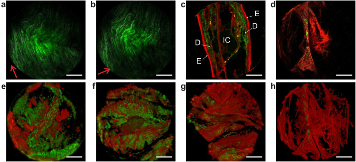
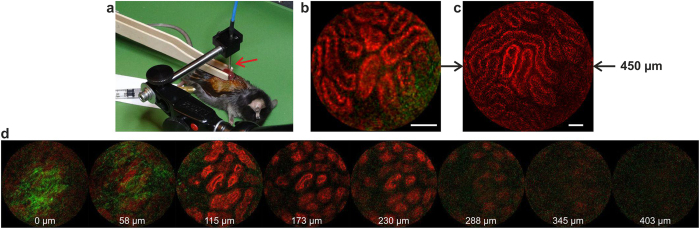
We present a two-photon microendoscope capable of in vivo label-free deep-tissue high-resolution fast imaging through a very long optical fiber. First, an advanced light-pulse spectro-temporal shaping device optimally precompensates for linear and nonlinear distortions occurring during propagation within the endoscopic fiber. This enables the delivery of sub-40-fs duration infrared excitation pulses at the output of 5 meters of fiber. Second, the endoscopic fiber is a custom-made double-clad polarization-maintaining photonic crystal fiber specifically designed to optimize the imaging resolution and the intrinsic luminescence backward collection. Third, a miniaturized fiber-scanner of 2.2 mm outer diameter allows simultaneous second harmonic generation (SHG) and two-photon excited autofluorescence (TPEF) imaging at 8 frames per second. This microendoscope's transverse and axial resolutions amount respectively to 0.8 μm and 12 μm, with a field-of-view as large as 450 μm. This microendoscope's unprecedented capabilities are validated during label-free imaging, ex vivo on various fixed human tissue samples, and in vivo on an anesthetized mouse kidney demonstrating an imaging penetration depth greater than 300 μm below the surface of the organ. The results reported in this manuscript confirm that nonlinear microendoscopy can become a valuable clinical tool for real-time in situ assessment of pathological states.
Figures


Similar articles
-
Reduction of self-phase modulation in double-clad photonic crystal fiber for nonlinear optical endoscopy.Opt Lett. 2009 Jan 15;34(2):148-50. doi: 10.1364/ol.34.000148. Opt Lett. 2009. PMID: 19148237
-
Compact and flexible raster scanning multiphoton endoscope capable of imaging unstained tissue.Proc Natl Acad Sci U S A. 2011 Oct 25;108(43):17598-603. doi: 10.1073/pnas.1114746108. Epub 2011 Oct 17. Proc Natl Acad Sci U S A. 2011. PMID: 22006303 Free PMC article.
-
Two-photon excited fluorescence lifetime measurements through a double-clad photonic crystal fiber for tissue micro-endoscopy.J Biophotonics. 2012 Jan;5(1):14-9. doi: 10.1002/jbio.201100070. Epub 2011 Nov 2. J Biophotonics. 2012. PMID: 22045513 Free PMC article.
-
Infrared multiphoton microscopy: subcellular-resolved deep tissue imaging.Curr Opin Biotechnol. 2009 Feb;20(1):54-62. doi: 10.1016/j.copbio.2009.02.008. Epub 2009 Mar 25. Curr Opin Biotechnol. 2009. PMID: 19324541 Review.
-
Fibre-optical microendoscopy.J Microsc. 2014 Apr;254(1):13-18. doi: 10.1111/jmi.12119. Epub 2014 Mar 5. J Microsc. 2014. PMID: 24593142 Review.
Cited by
-
Label-free highly multimodal nonlinear endoscope.Opt Express. 2022 Jul 4;30(14):25020-25033. doi: 10.1364/OE.462361. Opt Express. 2022. PMID: 36237042 Free PMC article.
-
Dual-modality endomicroscopy with co-registered fluorescence and phase contrast.Biomed Opt Express. 2016 Aug 10;7(9):3403-3411. doi: 10.1364/BOE.7.003403. eCollection 2016 Sep 1. Biomed Opt Express. 2016. PMID: 27699107 Free PMC article.
-
Spectro-temporal dispersion management of femtosecond pulses for fiber-optic two-photon endomicroscopy.Opt Express. 2018 Sep 3;26(18):22877-22893. doi: 10.1364/OE.26.022877. Opt Express. 2018. PMID: 30184945 Free PMC article.
-
Quantification of Collagen Organization after Nerve Repair.Plast Reconstr Surg Glob Open. 2017 Dec 11;5(12):e1586. doi: 10.1097/GOX.0000000000001586. eCollection 2017 Dec. Plast Reconstr Surg Glob Open. 2017. PMID: 29632766 Free PMC article.
-
Towards two-photon excited endogenous fluorescence lifetime imaging microendoscopy.Biomed Opt Express. 2017 Dec 8;9(1):142-156. doi: 10.1364/BOE.9.000142. eCollection 2018 Jan 1. Biomed Opt Express. 2017. PMID: 29359093 Free PMC article.
References
-
- Denk W., Strickler J. & Webb W. Two-photon laser scanning fluorescence microscopy. Science 248, 73–76 (1990). - PubMed
-
- Masters B. R. & So P. T. Biomedical applications of nonlinear optical microscopy in Handbook of biomedical nonlinear optical microscopy 705–844 (Oxford University, 2008).
-
- Wang C.-C. et al. Differentiation of normal and cancerous lung tissues by multiphoton imaging. J. Biomed. Opt. 14, 044034 (2009). - PubMed
Publication types
MeSH terms
LinkOut - more resources
Full Text Sources
Other Literature Sources
Medical
