. 2016 Mar;23(3):263-71.
doi: 10.1038/gt.2015.111. Epub 2015 Dec 16.

Mucopolysaccharidosis IIIB confers enhanced neonatal intracranial transduction by AAV8 but not by 5, 9 or rh10

Affiliations

Mucopolysaccharidosis IIIB confers enhanced neonatal intracranial transduction by AAV8 but not by 5, 9 or rh10

J A Gilkes et al. Gene Ther. 2016 Mar.

Abstract

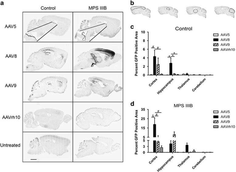
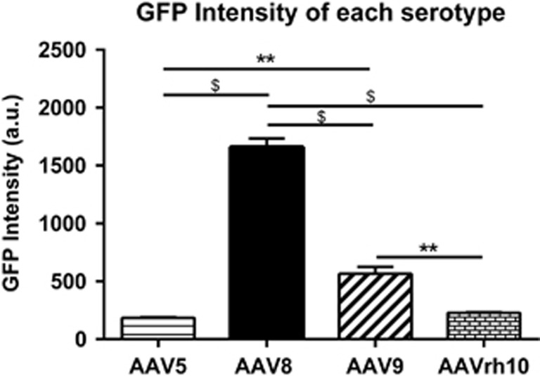
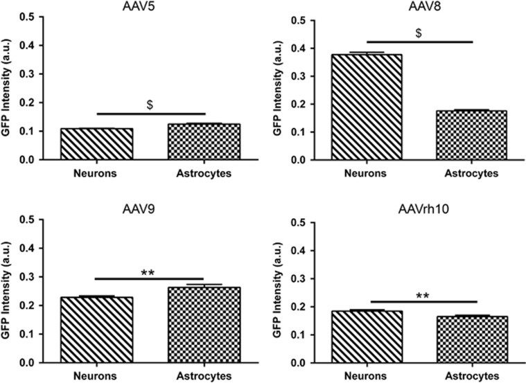
Sanfilippo syndrome type B (mucopolysaccharidosis IIIB, MPS IIIB) is a lysosomal storage disease resulting from deficiency of N-acetyl-glucosaminidase (NAGLU) activity. To determine the possible therapeutic utility of recombinant adeno-associated virus (rAAV) in early gene therapy-based interventions, we performed a comprehensive assessment of transduction and biodistribution profiles of four central nervous system (CNS) administered rAAV serotypes, -5, -8, -9 and -rh10. To simulate optimal earliest treatment of the disease, each rAAV serotype was injected into the CNS of neonatal MPS IIIB and control animals. We observed marked differences in biodistribution and transduction profiles between the serotypes and this differed in MPS IIIB compared with healthy control mice. Overall, in control mice, all serotypes performed comparably, although some differences were observed in certain focal areas. In MPS IIIB mice, AAV8 was more efficient than AAV5, -9 and -rh10 for gene delivery to most structures analyzed, including the cerebral cortex, hippocampus and thalamus. Noteworthy, the pattern of biodistribution within the CNS varied by serotype and genotype. Interestingly, AAV8 also produced the highest green fluorescent protein intensity levels compared with any other serotype and demonstrated improved transduction in NAGLU compared with control brains. Importantly, we also show leakage of AAV8, -9 and -rh10, but not AAV5, from CNS parenchyma to systemic organs. Overall, our data suggest that AAV8 represents the best therapeutic gene transfer vector for early intervention in MPS IIIB.


Figures

Figure 1
Figure 1
AAV serotype-dependent general CNS biodistribution. Following six site-directed vector administration (a), representative tissue sections of interest (b) were analyzed for percentage of area with GFP expression. Differences in combined percentage GFP area for all four sections were quantified for each serotype based on genotype (c), and rAAV-GFP expression was quantified for each selected brain section by serotype in 3-month-old control (d) and MPS IIIB littermates (e). Two-way ANOVA was used to assess differences within tissue section based on serotype, and one-way ANOVA was used to test individual differences between serotype, n=3–6/cohort *P<0.05; **P<0.01. Data represented as mean±s.e.m.
Figure 2
Figure 2
Mid-sagittal AAV serotype-dependent regional biodistribution. Mid-sagittal brain sections of control and MPS IIIB 3-month-old littermates were assessed for the presence of rAAV-GFP (dark areas, scale bar=2mm). Insets in AAV5 panels reflect transduced ependymal cells at × 20 magnification (a). Percentage area of GFP expression was assessed in the cortex, hippocampus, thalamus and cerebellum, outlined in (b) respectively of control (c) and MPS IIIB animals (d). All sagittal sections were scanned at × 20 magnification, and GFP expression was quantified using Aperio ImageScope algorythm. Two-way ANOVA was used to assess differences within each region based on serotype, and one-way ANOVA was used to test individual differences between serotype, n=3–6/cohort, *P<0.05, #P<0.001, $P<0.0001. Data represented as mean±s.e.m.
Figure 3
Figure 3
rAAVs exhibit differences in GFP production in MPS IIIB brain as indicated by resulting GFP intensity. Mid-sagittal sections of 3-month-old MPS IIIB animals were used to quantitate intensity of AAV-based GFP production. GFP intensities in the cortex, hippocampus, thalamus and cerebellum of each animal were summed. Data were analyzed by one-way ANOVA. **P<0.01, $P<0.0001. Data represented as mean±s.e.m., N=3/serotype.
Figure 4
Figure 4
All AAV serotypes transduce neurons and astrocytes but not microglia and oligodendrocytes in the cortex. Representative cortical images of AAV5, -8, -9 and -rh10 transduced MPS IIIB animals stained for neurons (NeuN), astrocytes (GFAP), microglia (Iba1) and oligodendrocytes (OSP). Arrows indicate areas of co-localization of each cell type and rAAV-GFP in the cortex of MPS IIIB animals. Images taken with × 40 objective lens, scale bar=60 μm.
Figure 5
Figure 5
Differences in GFP intensity of transduced cells are observed based on AAV serotype. GFP intensity of AAV transduced neurons and astrocytes was assessed in MPS IIIB animals at 3 months of age. Serotype-dependent differences in GFP intensity were determined using APERIO ScanScope FL Positive Pixel Count FL algorithm and quantitated using Student's t-test. N=3/serotype, **P<0.01, $P<0.0001. Data represented as mean±s.e.m.
Figure 6
Figure 6
CNS administered rAAVs also foster differential somatic transduction. Representative organs of 3-month-old control and MPS IIIB-treated animals were tested for somatic transduction of rAAV-GFP in the heart, liver, muscle, kidney and spleen. The presence of rAAV-GFP is indicated by black areas in each tissue section. All tissue sections were scanned at × 20 magnification, scale bar=100 μm.

References

    1. Yogalingam G, Hopwood JJ. Molecular genetics of mucopolysaccharidosis type IIIA and IIIB: diagnostic, clinical, and biological implications. Hum Mutat 2001; 18: 264–281. - PubMed
    1. Gilkes J, Patterson B, Heldermon C, Mucopolysaccharidosis III. Molecular genetics and genotype-phenotype correlations. OA Genet 2014; 2: 1.
    1. Li HH, Yu W-H, Rozengurt N, Zhao H-Z, Lyons KM, Anagnostaras S et al. Mouse model of Sanfilippo syndrome type B produced by targeted disruption of the gene encoding alpha-N-acetylglucosaminidase. Proc Natl Acad Sci USA 1999; 96: 14505–14510. - PMC - PubMed
    1. Heldermon CD, Hennig AK, Ohlemiller KK, Ogilvie JM, Herzog ED, Breidenbach et al. Development of sensory, motor and behavioral deficits in the murine model of Sanfilippo Syndrome Type B. PLoS ONE 2007; 2: e772. - PMC - PubMed
    1. Fu H, DiRosario J, Kang L, Muenzer J, McCarty DM. Restoration of central nervous system α-N-acetylglucosaminidase activity and therapeutic benefits in mucopolysaccharidosis IIIB mice by a single intracisternal recombinant adeno-associated viral type 2 vector delivery. J Gene Med 2010; 12: 624–633. - PubMed

Publication types