. 2016 Feb 1;143(3):473-82.
doi: 10.1242/dev.133843. Epub 2015 Dec 16.

Endocardial-to-mesenchymal transformation and mesenchymal cell colonization at the onset of human cardiac valve development

Affiliations

Endocardial-to-mesenchymal transformation and mesenchymal cell colonization at the onset of human cardiac valve development

Michael G Monaghan et al. Development. .

Abstract

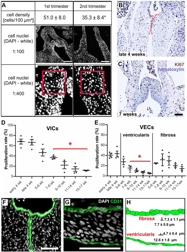
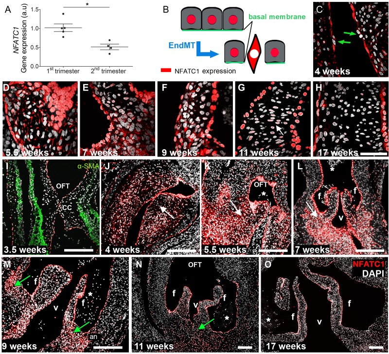
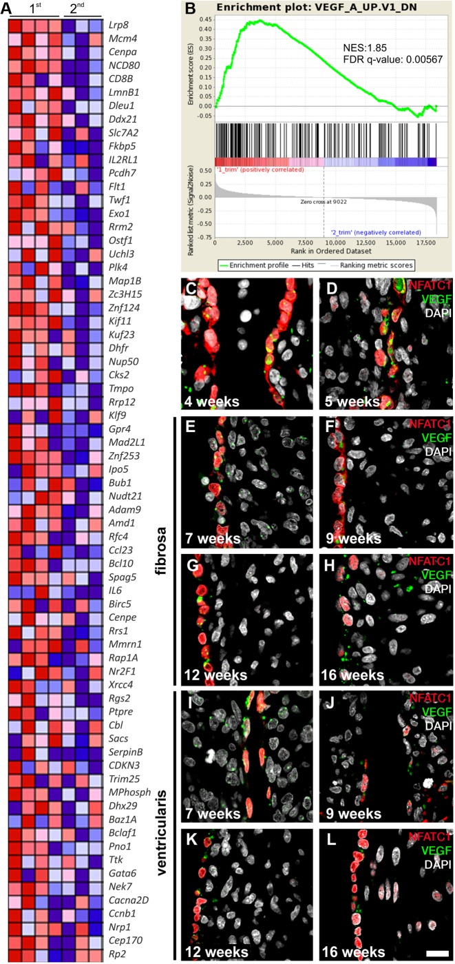
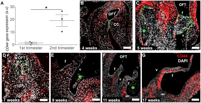
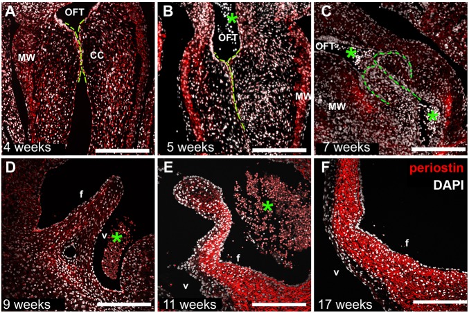
The elucidation of mechanisms in semilunar valve development might enable the development of new therapies for congenital heart disorders. Here, we found differences in proliferation-associated genes and genes repressed by VEGF between human semilunar valve leaflets from first and second trimester hearts. The proliferation of valve interstitial cells and ventricular valve endothelial cells (VECs) and cellular density declined from the first to the second trimester. Cytoplasmic expression of NFATC1 was detected in VECs (4 weeks) and, later, cells in the leaflet/annulus junction mesenchyme expressing inactive NFATC1 (5.5-9 weeks) were detected, indicative of endocardial-to-mesenchymal transformation (EndMT) in valvulogenesis. At this leaflet/annulus junction, CD44(+) cells clustered during elongation (11 weeks), extending toward the tip along the fibrosal layer in second trimester leaflets. Differing patterns of maturation in the fibrosa and ventricularis were detected via increased fibrosal periostin content, which tracked the presence of the CD44(+) cells in the second trimester. We revealed that spatiotemporal NFATC1 expression actively regulates EndMT during human valvulogenesis, as early as 4 weeks. Additionally, CD44(+) cells play a role in leaflet maturation toward the trilaminar structure, possibly via migration of VECs undergoing EndMT, which subsequently ascend from the leaflet/annulus junction.

Keywords: EndMT; Extracellular matrix; Heart; NFATc-1; Periostin; Semilunar valves.


Conflict of interest statement

Competing interests

The authors declare no competing or financial interests.

Figures

Fig. 1.
Fig. 1.
Cell density and proliferation decrease significantly from the first to the second trimester of leaflet development and defined VEC morphologies are visible as early as 4 weeks. (A) DAPI staining of human developing cardiac valves shows the different cell densities of the leaflets. The red square is 0.01 mm2. *P<0.001. (B,C) Proliferating KI67+ VICs (brown) are randomly distributed throughout the fetal cushions and leaflets during developmental stages. (B) Fetal semilunar valve cushions at late 4 weeks, and (C) leaflets at 7 weeks of development. Red lines highlight the semilunar cushions. (D,E) Proliferation rate of VICs and VECs on the ventricularis is significantly decreased beginning at 7-8 weeks of development when compared with late 4 weeks. *P<0.05. (F,G) CD31+ VECs in developing valves (green) show characteristically distinct morphologies: VECs with a cuboidal morphology line the fibrosa layer, whereas VECs facing the ventricles are elongated and flattened. (H) Schematic of cell morphologies. VECs of the fibrosa are significantly shorter than VECs of the ventricularis. VEC, ventricular valve endothelial cell; VIC, valvular interstitial cell. Scale bars: 200 µm in A; 100 µm in C,F,G.
Fig. 2.
Fig. 2.
The spatiotemporal patterns of NFATC1 gene and protein expression. (A) Gene expression of NFATC1 in first and second trimester semilunar valve leaflets (*P<0.006, n=4). (B) Schematic of spatial NFATC1 expression during EndMT. Active NFATC1 (red), maintaining the endothelial phenotype, is located in the cell nucleus. Although inactive, NFATC1 is expressed in the cytosol. (C-O) Immunofluorescence analyses of NFATC1 protein (red) expression patterns at the indicated stages. DAPI is in white. (C) The green arrows indicate cells appearing to undergo EndMT. (G) White arrow points to cells in the mesenchyme, where NFATC1 expression intensities in the nucleus are significantly decreased compared with endocardial cells. (I-O) During a specific time window between weeks 4 and 8, NFATC1 is also present in the cytosol (white arrows) of cushion mesenchymal cells (J-L). Between week 9 (M) and 11 (N), NFATC1 is expressed in the nucleus (green arrows) of mesenchymal cells at the leaflet annulus. In second trimester leaflets (O), nuclear NFATC1 is predominately found in VECs. an, leaflet annulus; OFT, outflow tract; f, fibrosa; v, ventricularis; CC, cardiac cushions; asterisks, erythrocytes. Scale bars: 50 µm in C-H; 200 µm in I-O.
Fig. 3.
Fig. 3.
Increased expression of VEGF targets in the first trimester. (A) Heat map representing the relative expression and fold enrichment of genes repressed by VEGF (VEGFA) in the first (n=4) and second (n=3) trimester in OFT leaflets (created using Molecular Signatures Database v5.0). See B for color key. (B) GSEA of enrichment of VEGF repression from leaflets obtained from the first compared with the second trimester. The false discovery rate (FDR) q-value and normalized enrichment score (NES) are shown. (C-L) Immunofluorescence imaging of NFATC1 (red) and VEGF (green) protein expression patterns in early endocardial cushions and elongated leaflets. DAPI is in white. At the early cushion stage (C,D), fibrosa and ventricularis are not yet identifiable. E-H, the fibrosa layer; I-L, the ventricularis. Scale bar: 50 µm.
Fig. 4.
Fig. 4.
Increased expression of CD44 from the first to the second trimester and the spatiotemporal pattern of CD44 protein expression. CD44 gene (A; *P<0.05, n=3) and protein (B-G) expression analyses reveal the distinct temporal and spatial patterns of CD44+ cells (red) during human semilunar valvulogenesis. DAPI is in white. The green lines highlight the semilunar cushions. OFT, outflow tract; f, fibrosa; v, ventricular side; CC, cardiac cushions; an, leaflet annulus; asterisks, erythrocytes. Scale bars: 200 µm in B-E; 400 µm in F,G.
Fig. 5.
Fig. 5.
Periostin expression accumulates strongly in the fibrosal layer of the developing leaflet in the second trimester. Temporal and spatial distribution of periostin (red) in human first (A-E) and second (F) trimester semilunar leaflets. DAPI is in white. The green lines highlight the semilunar cushions. OFT, outflow tract; asterisks, erythrocytes; f, fibrosa; v, ventricularis; CC, cardiac cushions; MW, myocardial wall. Scale bars: 200 µm in A-D; 400 µm in E,F.

References

    1. Aikawa E., Whittaker P., Farber M., Mendelson K., Padera R. F., Aikawa M. and Schoen F. J. (2006). Human semilunar cardiac valve remodeling by activated cells from fetus to adult: implications for postnatal adaptation, pathology, and tissue engineering. Circulation 113, 1344-1352. 10.1161/CIRCULATIONAHA.105.591768 - DOI - PubMed
    1. Armstrong E. J. and Bischoff J. (2004). Heart valve development: endothelial cell signaling and differentiation. Circ. Res. 95, 459-470. 10.1161/01.RES.0000141146.95728.da - DOI - PMC - PubMed
    1. Butcher J. T. and Markwald R. R. (2007). Valvulogenesis: the moving target. Philos. Trans. R. Soc. Lond. B Biol. Sci. 362, 1489-1503. 10.1098/rstb.2007.2130 - DOI - PMC - PubMed
    1. Carthy J. M., Boroomand S. and McManus B. M. (2012). Versican and CD44 in in vitro valvular interstitial cell injury and repair. Cardiovasc. Pathol. 21, 74-82. 10.1016/j.carpath.2011.03.003 - DOI - PubMed
    1. Chang C.-P., Neilson J. R., Bayle J. H., Gestwicki J. E., Kuo A., Stankunas K., Graef I. A. and Crabtree G. R. (2004). A field of myocardial-endocardial NFAT signaling underlies heart valve morphogenesis. Cell 118, 649-663. 10.1016/j.cell.2004.08.010 - DOI - PubMed

Publication types

MeSH terms

Substances