. 2015 Dec 17:6:10215.
doi: 10.1038/ncomms10215.

Cdk1 orders mitotic events through coordination of a chromosome-associated phosphatase switch

Affiliations

Cdk1 orders mitotic events through coordination of a chromosome-associated phosphatase switch

Junbin Qian et al. Nat Commun. .

Abstract

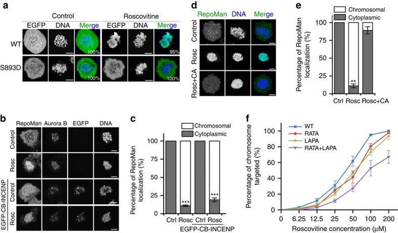
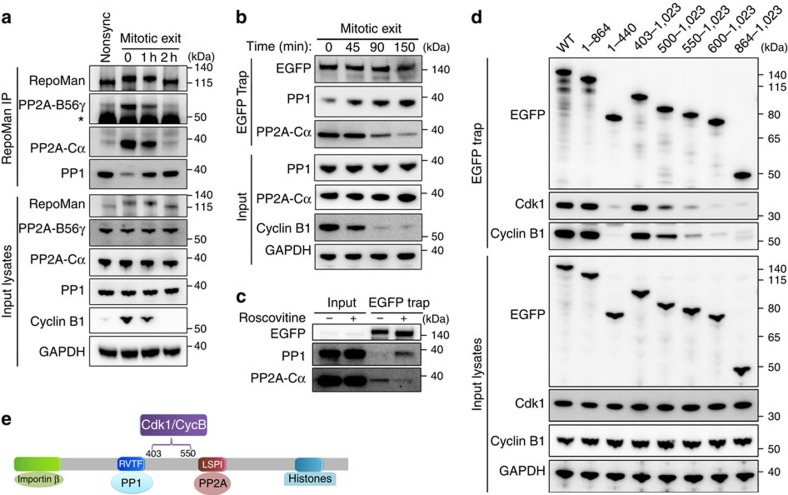
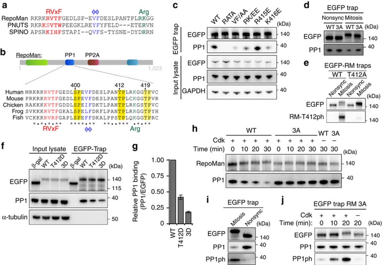
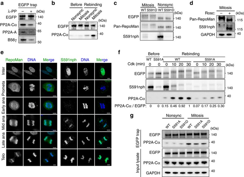
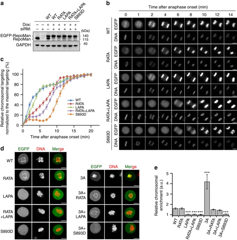
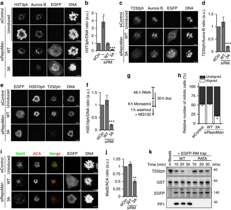
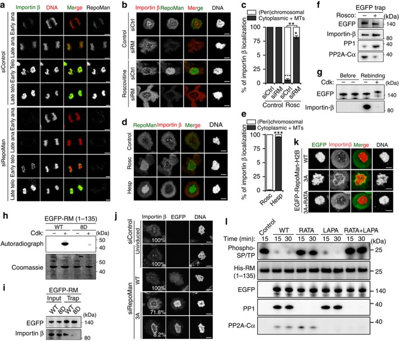
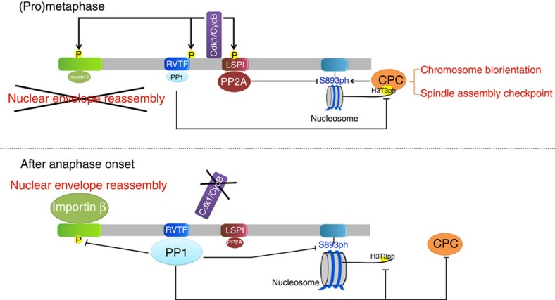
RepoMan is a scaffold for signalling by mitotic phosphatases at the chromosomes. During (pro)metaphase, RepoMan-associated protein phosphatases PP1 and PP2A-B56 regulate the chromosome targeting of Aurora-B kinase and RepoMan, respectively. Here we show that this task division is critically dependent on the phosphorylation of RepoMan by protein kinase Cyclin-dependent kinase 1 (Cdk1), which reduces the binding of PP1 but facilitates the recruitment of PP2A-B56. The inactivation of Cdk1 in early anaphase reverses this phosphatase switch, resulting in the accumulation of PP1-RepoMan to a level that is sufficient to catalyse its own chromosome targeting in a PP2A-independent and irreversible manner. Bulk-targeted PP1-RepoMan also inactivates Aurora B and initiates nuclear-envelope reassembly through dephosphorylation-mediated recruitment of Importin β. Bypassing the Cdk1 regulation of PP1-RepoMan causes the premature dephosphorylation of its mitotic-exit substrates in prometaphase. Hence, the regulation of RepoMan-associated phosphatases by Cdk1 is essential for the timely dephosphorylation of their mitotic substrates.


Figures

Figure 1
Figure 1. Cdk1 opposes the chromosome targeting of RepoMan.
(a) Nocodazole-arrested U2OS cells transiently expressing EGFP-tagged RepoMan-WT or RepoMan-S893D were treated with dimethyl sulfoxide (DMSO) (control) or Roscovitine (100 μM) for 30 min before fixation and visualization of EGFP and DNA. The numbers in the figures refer to the percentage of cells where the shown phenotype occurred. Scale bars, 5 μm. (b) Confocal images of Nocodazole-arrested U2OS cells, in the absence or presence of Roscovitine (Rosc) and transiently expressed EGFP-tagged CENP-B (CB) fused to INCENP. The fixed cells were stained for endogenous RepoMan, Aurora B and DNA. Scale bars, 5 μm. (c) Quantification of RepoMan localization in b. The graphs show mean percentages±s.e.m. from three independent experiments (≥73 cells for each condition per experiment). ***P<0.001 in paired t-test, compared with control (Ctrl). (d) Nocodazole-arrested U2OS cells were treated with DMSO (control) or 100 μM Roscovitine for 30 min, as such or in combination with 20 nM Calyculin A (CA). The fixed cells were stained for endogenous RepoMan and DNA. Scale bars, 5 μm. (e) Quantification of EGFP-RepoMan localization in d. Graphs show mean percentages±s.e.m. from three independent experiments (≥74 cells for each condition per experiment). **P<0.01 in paired t-test, compared with control (Ctrl). (f) Nocodazole-arrested U2OS cells expressing the indicated EGFP-tagged RepoMan variants were incubated for 30 min with different concentrations of Roscovitine before fixation. DNA was stained with DAPI. The figure shows the percentage of cells where the EGFP-fusion was chromosome-associated. The results are expressed as means±s.e.m. from three independent experiments (≥103 cells per data point).
Figure 2
Figure 2. Cdk1 regulates the association of PP1 and PP2A-B56 with RepoMan.
(a) Nocodazole-arrested prometaphase U2OS cells were collected by shake-off and then released into fresh medium for different time points to synchronize the cells for mitotic exit. Immunoprecipitates (IP) of endogenous RepoMan were prepared from lysates of these synchronized cells as well as from non-synchronized cells. The IPs were analysed by immunoblotting with the indicated antibodies. Asterisk denotes IgG-heavy chain. (b) U2OS cells were transfected with EGFP-RepoMan-WT and arrested/released as in a. EGFP traps of cell lysates were analysed by immunoblotting. (c) Nocodazole-arrested U2OS cells were collected by shake-off and then treated with or without 100 μM Roscovitine for 30 min. EGFP traps from cell lysates were analysed by immunoblotting. (d) EGFP traps of lysates from Nocodazole-arrested HEK293T cells expressing EGFP fusions of the indicated RepoMan fragments were processed for immunoblotting with the indicated antibodies. (e) Map of the protein-binding sites of RepoMan.
Figure 3
Figure 3. Cdk1 reduces the activity of PP1-RepoMan.
(a) Sequence alignment of the PP1-interaction domains of RepoMan, PNUTS and Spinophilin. (b) Conservation of the PP1-interaction domain of RepoMan in vertebrates. (c) EGFP traps from HEK293T cells expressing the indicated EGFP-RepoMan mutants were analysed by immunoblotting for the binding of PP1. (d) U2OS cells were transfected with EGFP-tagged RepoMan-WT or RepoMan-3A. EGFP traps from non-synchronized (Nonsync) or Nocodazole-arrested (Mitosis) U2OS cell lysates were examined for associated PP1. (e) U2OS cells were transfected with EGFP-tagged RepoMan-WT. EGFP traps from Nonsync or Nocodazole-arrested (Mitosis) U2OS cell lysates were examined for EGFP and RepoMan-T412ph antibody. (f) EGFP traps from non-synchronized HEK293T cells expressing EGFP-tagged β-galactosidase (β-gal), RepoMan-WT, RepoMan-T412D or RepoMan-3D were analysed by immunoblotting for associated PP1. (g) The PP1/EGFP ratio was quantified by densitometric analysis. The data represent means±s.e.m. from three independent experiments. (h) EGFP traps from non-synchronized HEK293T cells expressing EGFP-tagged RepoMan-WT or RepoMan-3A were phosphorylated in vitro with recombinant Cdk2/Cyclin A. Unbound PP1 was washed away before the addition of SDS sample buffer to the traps and immunoblotting for PP1. (i) Phosphorylation of the C-terminal Cdk site of PP1 (PP1ph) was examined by immunoblotting of EGFP traps from U2OS cells expressing EGFP-RepoMan-WT in Nocodazole-arrested (Mitosis) or non-synchronized (Nonsync) U2OS cells with a phospho-epitope specific antibody. (j) EGFP traps from non-synchronized HEK293T cells expressing EGFP-RepoMan-3A (RM 3A) were phosphorylated with Cdk2/Cyclin A. The unbound fractions were washed away before the addition of SDS sample buffer to the traps and immunoblotting for PP1 and PP1ph.
Figure 4
Figure 4. Cdk1 promotes the binding of PP2A-B56 to RepoMan.
(a) Traps of EGFP-RepoMan-WT from Nocodazole-arrested U2OS cells were treated with or without λ-phosphatase for 30 min at 30 °C. The proteins released from the traps were washed away before the addition of SDS-sample buffer to the traps and immunoblot analysis for the presence of associated PP2A-B56 subunits. (b) Traps of EGFP-RepoMan-WT from non-synchronized or Nocodazole-arrested U2OS cells were washed with 1.5 M NaCl to remove associated PP2A (Before). Subsequently, the traps were added to freshly prepared mitotic lysates and asssociated PP2A was quantified by immunoblotting of the pelleted traps (Rebinding). (c) Cell lysates were prepared from Nocodazole-arrested (Mitosis) or non-synchronized (Nonsync) U2OS cells expressing EGFP-tagged RepoMan-WT or RepoMan-S591D. The EGFP traps were analysed by immunoblotting for EGFP, RepoMan (pan-RepoMan) or the phosphorylation of RepoMan at S591 (S591nph). (d) Nocodazole-arrested U2OS cells were treated with or without 100 μM Roscovitine for 1 h. The phosphorylation of S591 was examined by immunoblotting with the S591nph antibody. (e) Immunofluorescence staining of unperturbed U2OS cells at different stages of the cell cycle with the pan-RepoMan or S591nph antibodies. Scale bars represent 5 μm. (f) EGFP traps from lysates of non-synchronized HEK293T cells expressing EGFP-tagged RepoMan-WT or RepoMan-S591A were washed with 1.5 M NaCl to remove associated PP2A. Subsequently, the traps were phosphorylated with Cdk2 for the indicated time points and added to freshly prepared mitotic cell lysates from HeLa cells to examine PP2A binding to the pelleted traps. Phosphorylation of the fusions is suggested by mobility shifts and the loss of S591nph. (g) Traps of EGFP-tagged RepoMan-WT, RepoMan-S591A or RepoMan-S591D from non-synchronized (Nonsync) or Nocodazole-arrested (Mitosis) U2OS cells were analysed by immunoblotting for the binding of PP2A.
Figure 5
Figure 5. The chromosome targeting of RepoMan is regulated by both PP2A-B56 and PP1.
(a) One day after the knockdown (siRM) of endogenous RepoMan in HeLa Flp-in T-Rex cells, siRNA-resistant EGFP-RepoMan fusions were induced for 24 h with doxycycline (Dox). Endogenous RepoMan and the EGFP-fusions were visualized by immunoblotting with pan-RepoMan antibodies. (b) Same as in a but the cells were subjected to time-lapse imaging by confocal microscopy after release from a 6 h-Monastrol arrest. The panel shows cells at different time points after the initiation of anaphase. DNA was stained with Hoechst 33342. Scale bars, 5 μm. (c) Quantification of the chromosome targeting (EGFP/DNA ratio) of the EGFP-RepoMan fusions as shown in (b). The means±s.e.m. of at least 11 cells are shown in each condition. Data were normalized to the maximal ratio of EGFP/DNA in each condition. (d) Live imaging of Nocodazole-arrested U2OS cells after expression of EGFP-tagged RepoMan or its phosphatase/histone-binding mutants. The figure shows the localization of DNA and the EGFP fusions. Scale bars, 5 μm. (e) Quantification of the ratio of EGFP signal on chromosome versus cytoplasm, as shown in d. The means±s.e.m. of at least 12 cells were analysed in each condition. ***P<0.001 in paired t-test, as compared with WT.
Figure 6
Figure 6. The inhibition of PP1-RepoMan maintains Aurora B activity in prometaphase.
(a) HeLa Flp-in T-Rex cells were induced to express siRNA-resistant EGFP-tagged RepoMan-WT or RepoMan-3A following the knockdown of endogenous RepoMan. Monastrol-arrested mitotic cells were fixed before staining. Scale bars, 5 μm. (b) Quantification of the H3T3ph/DNA ratio in a. The data represent means±s.e.m. for three independent experiments (≥11 cells per condition in each experiment). (c) Cells were treated as in a but stained for Aurora B, Aurora-B -T232ph and DNA. Scale bars, 5 μm. (d) Quantification of the T232ph/Aurora-B ratio in c. The data represent means±s.e.m. for three independent experiments (≥11 cells per condition in each experiment) with paired t-test, as compared with siCtrl. (e) Cells were treated as in a but stained for H3S10ph, Aurora-B and DNA. Scale bars, 5 μm. (f) Quantification of the H3S10ph/DNA ratio in e. The data represent means±s.e.m. for three independent experiments (≥10 cells per condition in each experiment). (g) Cell-treatment scheme for chromosome alignment assays of HeLa Flp-in T-Rex cells, before and after induction of EGFP-tagged RepoMan-WT or RepoMan-3A, and following the knockdown of endogenous RepoMan. (h) In cells treated as explained in g, the percentage of cells with a well-aligned metaphase plate or unaligned chromosomes were plotted as mean percentages±s.e.m. for three independent experiments (≥121 cells per condition in each experiment). (i) Cells were treated as in a but stained for Mad2, ACA and DNA. (j) Quantification of relative signal of centromeric Mad2 in i. Data were normalized to the indicated ratio as means±s.e.m. for three independent experiments (≥8 centromeres per cell, ≥10 cells per condition in each experiment). (k) Traps of EGFP-fusions with RepoMan-WT or RepoMan-RATA from non-synchronized HEK293T cells were used as a phosphatase source to dephosphorylate recombinant GST-Aurora B/INCENP. The figure shows different time points of incubation with the traps analysed by immunoblotting. *P<0.05; **P<0.01; ***P<0.001, with paired t-test as compared with siCtrl.
Figure 7
Figure 7. Cdk1 and PP1 antagonistically regulate the RepoMan/Importin β interaction.
(a) U2OS cells transfected with control or RepoMan siRNA were fixed and stained after pre-extraction with 0.2% Triton X-100. Scale bars, 5 μm. (b) Nocodazole-arrested U2OS cells transfected with control or RepoMan siRNA were treated with DMSO or 100 μM Roscovitine for 30 min and subsequently stained. Scale bars, 5 μm. (c) Quantification for Importin-β localization as shown in b. The data are represented as mean percentages±s.e.m. for three independent experiments (≥66 cells per condition in each experiment). *P<0.05; **P<0.01; ***P<0.001 with paired t-test, as compared with siCtrl. (d) Nocodazole-arrested U2OS cells were treated with or without 100 μM Roscovitine (Rosc) or 200 nM Hesperadin (Hesp) for 30 min before staining. Scale bars, 5 μm. (e) Quantification for Importin-β localization as shown in d. The data are represented as mean percentages±s.e.m. for four independent experiments (≥76 cells per condition in each experiment). ***P<0.001 with paired t-test, as compared with Rosc. (f) Nocodazole-arrested U2OS cells expressing EGFP-RepoMan-WT were treated with DMSO or Roscovitine for 30 min. EGFP traps from the cell lysates were analysed by immunoblotting with the indicated antibodies. (g) EGFP traps from non-synchronized HEK293T cells expressing EGFP-RepoMan-WT were treated with 1.5 M NaCl, subjected to in vitro phosphorylation by Cdk2, and subsequently examined for Importin-β binding before or after incubation with mitotic cell lysates (Rebinding). (h) EGFP traps from HEK293T cells expressing EGFP-tagged RepoMan-(1–135)-WT or RepoMan-(1–135)-8D were radioactively phosphorylated with Cdk2 and analysed by autoradiography and Coomassie staining. (i) EGFP traps from HEK293T cells expressing EGFP-tagged RepoMan-WT or RepoMan-8D were analysed by immunoblotting for the binding of Importin β. (j) Nocodazole-arrested HeLa Flp-in T-Rex cells were transfected with control or RepoMan siRNAs, and induced to express EGFP-tagged RepoMan-WT or RepoMan-3A before staining. The numbers in the figures refer to the percentage of cells where the shown phenotype occurred. Scale bars, 5 μm. (k) Nocodazole-arrested Hela cells expressing H2B fusions of RepoMan-WT, RepoMan-3A and RepoMan-3A+RATA. Scale bars, 5 μm. (l) EGFP traps of the indicated RepoMan variants from non-synchronized HEK293T cells were used as a phosphatase source to dephosphorylate Cdk2-phosphorylated His-tagged RepoMan-(1–135).
Figure 8
Figure 8. Model of the Cdk1-mediated coordination of RepoMan-associated phosphatases.
During (pro)metaphase, Cdk1 promotes Aurora-B activation and prevents nuclear-envelope reassembly through phosphorylation of RepoMan at multiple sites, which reduces the binding of PP1 and Importin β. In contrast, the Cdk1-mediated phosphorylation of RepoMan at S591 enhances the recruitment of PP2A, which contributes to the dynamic chromosome targeting of RepoMan in (pro)metaphase and the centromeric localization of Aurora B. After anaphase onset, Cdk1 inactivation leads to the dephosphorylation of RepoMan, associated with the loss of PP2A and the bulk recruitment of PP1-RepoMan to the chromosomes. PP1-RepoMan then inactivates Aurora B and promotes nuclear-envelope reassembly through dephosphorylation of Histone H3 at T3, Aurora B at T232 and RepoMan at S893 and the Importin-β-binding domain.

References

    1. Malumbres M. Cyclin-dependent kinases. Genome Biol. 15, 122 (2014). - PMC - PubMed
    1. Tsukahara T., Tanno Y. & Watanabe Y. Phosphorylation of the CPC by Cdk1 promotes chromosome bi-orientation. Nature 467, 719–723 (2010). - PubMed
    1. Ghenoiu C., Wheelock M. & Funabiki H. Autoinhibition and polo-dependent multisite phosphorylation restrict activity of the histone H3 kinase haspin to mitosis. Mol. Cell 52, 734–745 (2013). - PMC - PubMed
    1. Zhou L., Tian X., Zhu C., Wang F. & Higgins J. M. Polo-like kinase-1 triggers Histone phosphorylation by Haspin in mitosis. EMBO Rep. 15, 273–281 (2014). - PMC - PubMed
    1. Bollen M., Gerlich D. W. & Lesage B. Mitotic phosphatases: from entry guards to exit guides. Trends Cell Biol. 19, 531–541 (2009). - PubMed

Publication types

MeSH terms

LinkOut - more resources