

Decreased PCSK9 expression in human hepatocellular carcinoma
- PMID: 26674961
- PMCID: PMC4682218
- DOI: 10.1186/s12876-015-0371-6
Decreased PCSK9 expression in human hepatocellular carcinoma
Abstract
Background: The management of hepatocellular carcinoma (HCC) is limited by the lack of adequate screening biomarkers and chemotherapy. In response, there has been much interest in tumor metabolism as a therapeutic target. PCSK9 stimulates internalization of the LDL-receptor, decreases cholesterol uptake into hepatocytes and affects liver regeneration. Thus, we investigated whether PCSK9 expression is altered in HCC, influencing its ability to harness cholesterol metabolism.
Methods: Thirty-nine patients undergoing partial hepatectomy or liver transplantation for HCC were consented for use of HCC tissue to construct a tissue microarray (TMA). The TMA was immunostained for PCSK9. Imagescope software was used to objectively determine staining, and assess for pathological and clinical correlations. PCSK9 and LDL receptor mRNA levels in flash-frozen HCC and adjacent liver tissue were determined by quantitative RT-PCR. Serum PCSK9 levels were determined by ELISA.
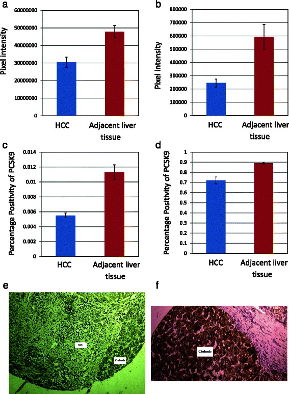
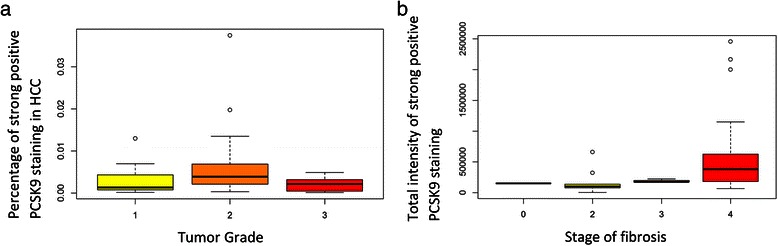
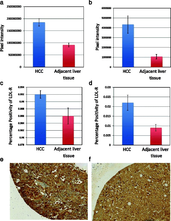
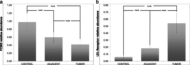
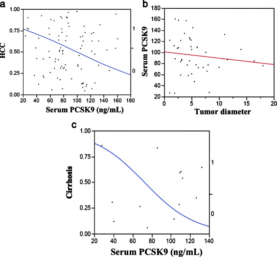
Results: By immunohistochemistry, there was significantly lower expression of PCSK9 in HCC as compared to adjacent cirrhosis (p-value < 0.0001, wilcoxon signed-rank test). Significantly greater staining of PCSK9 was present in cirrhosis compared to HCC (p value <0.0001), and positivity (percentage of positive cells) was significantly greater in cirrhosis compared to HCC (p-value < 0.0001). Conversely, significantly higher expression of LDL-R was present in HCC as compared to the adjacent cirrhosis (p-value < 0.0001). There was no significant correlation of PCSK9 staining with grade of tumor, but there were significant correlations between PCSK9 staining and stage of fibrosis, according to spearman correlation test. PCSK9 mRNA levels were relatively less abundant within HCC compared to adjacent liver tissue (p-value =0.08) and normal control tissue (p-value =0.02). In contrast, serum PCSK9 levels were significantly increased among patients with HCC compared to those with chronic liver disease without HCC (p-value =0.029). LDL receptor mRNA was consistantly greater in HCC when compared to normal control tissue (p-value = 0.06) and, in general, was significantly greater in HCC when compared to adjacent liver (p-value = 0.04).
Conclusions: The decreased expression of PCSK9 and conversely increased LDL-R expression in HCC suggests that HCC modulates its local microenvironment to enable a constant energy supply. Larger-scale studies should be conducted to determine whether PCSK9 could be a therapeutic target for HCC.
Figures
Similar articles
-
Serum proprotein convertase subtilisin/kexin type 9 and cell surface low-density lipoprotein receptor: evidence for a reciprocal regulation.Circulation. 2013 Jun 18;127(24):2403-13. doi: 10.1161/CIRCULATIONAHA.113.001592. Epub 2013 May 20. Circulation. 2013. PMID: 23690465 Free PMC article.
-
Serum proprotein convertase subtilisin/kexin type 9 concentration is not increased by plant stanol ester consumption in normo- to moderately hypercholesterolaemic non-obese subjects. The BLOOD FLOW intervention study.Clin Sci (Lond). 2015 Sep;129(5):439-46. doi: 10.1042/CS20150193. Epub 2015 Apr 10. Clin Sci (Lond). 2015. PMID: 25857271 Clinical Trial.
-
Plasma proprotein convertase subtilisin/kexin type 9 is associated with Lp(a) in type 2 diabetic patients.J Diabetes Complications. 2015 Nov-Dec;29(8):1165-70. doi: 10.1016/j.jdiacomp.2015.08.003. Epub 2015 Aug 10. J Diabetes Complications. 2015. PMID: 26412029
-
On the function and homeostasis of PCSK9: reciprocal interaction with LDLR and additional lipid effects.Atherosclerosis. 2015 Feb;238(2):264-70. doi: 10.1016/j.atherosclerosis.2014.12.017. Epub 2014 Dec 17. Atherosclerosis. 2015. PMID: 25544176 Free PMC article. Review.
-
Hypobetalipoproteinemia and abetalipoproteinemia.Curr Opin Lipidol. 2014 Jun;25(3):161-8. doi: 10.1097/MOL.0000000000000072. Curr Opin Lipidol. 2014. PMID: 24751931 Free PMC article. Review.
Cited by
-
Antitumor activity and molecular mechanism of proprotein convertase subtilisin/kexin type 9 (PCSK9) inhibition.Naunyn Schmiedebergs Arch Pharmacol. 2022 Jun;395(6):643-658. doi: 10.1007/s00210-022-02200-y. Epub 2022 Mar 21. Naunyn Schmiedebergs Arch Pharmacol. 2022. PMID: 35307759 Review.
-
Rewiring Lipid Metabolism by Targeting PCSK9 and HMGCR to Treat Liver Cancer.Cancers (Basel). 2022 Dec 20;15(1):3. doi: 10.3390/cancers15010003. Cancers (Basel). 2022. PMID: 36612001 Free PMC article.
-
Accumulation of cholesterol, triglycerides and ceramides in hepatocellular carcinomas of diethylnitrosamine injected mice.Lipids Health Dis. 2021 Oct 10;20(1):135. doi: 10.1186/s12944-021-01567-w. Lipids Health Dis. 2021. PMID: 34629057 Free PMC article.
-
Cholesterol metabolism in tumor microenvironment: cancer hallmarks and therapeutic opportunities.Int J Biol Sci. 2024 Mar 17;20(6):2044-2071. doi: 10.7150/ijbs.92274. eCollection 2024. Int J Biol Sci. 2024. PMID: 38617549 Free PMC article. Review.
-
Drug Repurposing Flubendazole to Suppress Tumorigenicity via PCSK9-dependent Inhibition and Potentiate Lenvatinib Therapy for Hepatocellular Carcinoma.Int J Biol Sci. 2023 Apr 23;19(7):2270-2288. doi: 10.7150/ijbs.81415. eCollection 2023. Int J Biol Sci. 2023. PMID: 37151886 Free PMC article.
References
Publication types
MeSH terms
Substances
LinkOut - more resources
Full Text Sources
Other Literature Sources
Medical
Miscellaneous
