

Noninvasive diagnostics of mitochondrial disorders in isolated lymphocytes with high resolution respirometry
- PMID: 26675066
- PMCID: PMC4633944
- DOI: 10.1016/j.bbacli.2014.09.003
Noninvasive diagnostics of mitochondrial disorders in isolated lymphocytes with high resolution respirometry
Abstract
Background: Mitochondrial diseases belong to the most severe inherited metabolic disorders affecting pediatric population. Despite detailed knowledge of mtDNA mutations and progress in identification of affected nuclear genes, diagnostics of a substantial part of mitochondrial diseases relies on clinical symptoms and biochemical data from muscle biopsies and cultured fibroblasts.
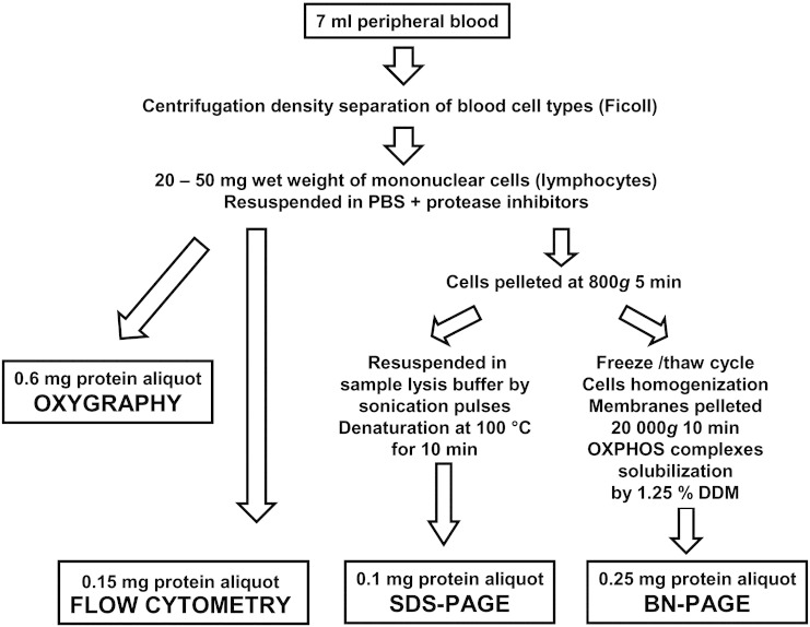
Methods: To investigate manifestation of oxidative phosphorylation defects in isolated lymphocytes, digitonin-permeabilized cells from 48 children were analyzed by high resolution respirometry, cytofluorometric detection of mitochondrial membrane potential and immunodetection of respiratory chain proteins with SDS and Blue Native electrophoreses.
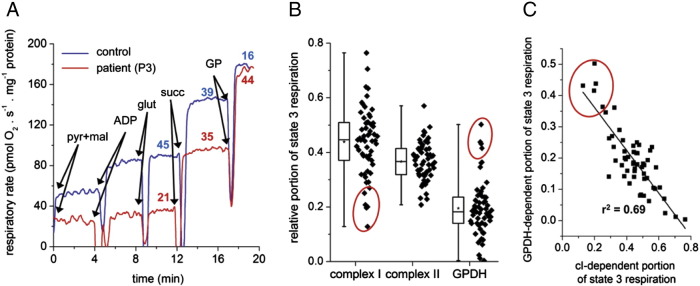
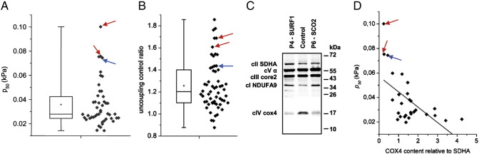
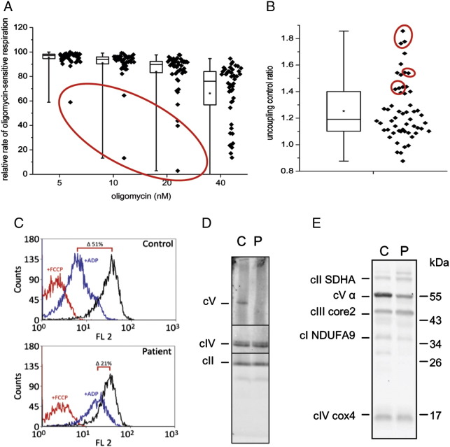
Results: Evaluation of individual respiratory complex activities, ATP synthesis, kinetic parameters of mitochondrial respiratory chain and the content and subunit composition of respiratory chain complexes enabled detection of inborn defects of respiratory complexes I, IV and V within 2 days. Low respiration with NADH-dependent substrates and increased respiration with glycerol-3-phosphate revealed complex I defects; changes in p 50 for oxygen and elevated uncoupling control ratio pointed to complex IV deficiency due to SURF1 or SCO2 mutation; high oligomycin sensitivity of state 3-ADP respiration, upregulated mitochondrial membrane potential and low content of complex V were found in lymphocytes with ATP synthase deficiency due to TMEM70 mutations.
Conclusion: Based on our results, we propose the best biochemical parameters predictive for defects of respiratory complexes I, IV and V manifesting in peripheral blood lymphocytes.
General significance: The noninvasiveness, reliability and speed of an approach utilizing novel biochemical criteria demonstrate the high potential of isolated lymphocytes for diagnostics of oxidative phosphorylation disorders in pediatric patients.
Keywords: AA, antimycin A; BNE, Blue Native PAGE; COX, cytochrome c oxidase; Diagnostics; FCCP, carbonyl cyanide 4-(trifluoromethoxy)phenylhydrazone; GP, glycerol-3-phosphate; GPDH, mitochondrial FAD-dependent glycerophosphate dehydrogenase; Lymphocytes; Mitochondrial diseases; OXPHOS, oxidative phosphorylation; Oxidative phosphorylation; PAGE, polyacrylamide gel electrophoresis; Respirometry; TMPD, tetramethylphenylenediamine; TMRM, tetramethylrhodamine methyl ester; cI–cV, respiratory chain complexes I–V; s3, state 3-ADP; s3u, state 3-uncoupled; s4o, state 4-oligomycin; ΔΨm, mitochondrial membrane potential.
Figures
Similar articles
-
High resolution respirometry analysis of polyethylenimine-mediated mitochondrial energy crisis and cellular stress: Mitochondrial proton leak and inhibition of the electron transport system.Biochim Biophys Acta. 2013 Oct;1827(10):1213-25. doi: 10.1016/j.bbabio.2013.07.001. Epub 2013 Jul 11. Biochim Biophys Acta. 2013. PMID: 23850549
-
Availability of the key metabolic substrates dictates the respiratory response of cancer cells to the mitochondrial uncoupling.Biochim Biophys Acta. 2014 Jan;1837(1):51-62. doi: 10.1016/j.bbabio.2013.07.008. Epub 2013 Jul 23. Biochim Biophys Acta. 2014. PMID: 23891695
-
The branched mitochondrial respiratory chain from Debaryomyces hansenii: components and supramolecular organization.Biochim Biophys Acta. 2014 Jan;1837(1):73-84. doi: 10.1016/j.bbabio.2013.07.011. Epub 2013 Aug 7. Biochim Biophys Acta. 2014. PMID: 23933018
-
Mitochondria and diabetes. Genetic, biochemical, and clinical implications of the cellular energy circuit.Diabetes. 1996 Feb;45(2):113-26. doi: 10.2337/diab.45.2.113. Diabetes. 1996. PMID: 8549853 Review.
-
Mitochondrial medicine--molecular pathology of defective oxidative phosphorylation.Ann Clin Lab Sci. 2001 Jan;31(1):25-67. Ann Clin Lab Sci. 2001. PMID: 11314862 Review.
Cited by
-
Utilization of Human Samples for Assessment of Mitochondrial Bioenergetics: Gold Standards, Limitations, and Future Perspectives.Life (Basel). 2021 Sep 10;11(9):949. doi: 10.3390/life11090949. Life (Basel). 2021. PMID: 34575097 Free PMC article. Review.
-
Coenzyme Q10: A Biomarker in the Differential Diagnosis of Parkinsonian Syndromes.Antioxidants (Basel). 2023 Dec 12;12(12):2104. doi: 10.3390/antiox12122104. Antioxidants (Basel). 2023. PMID: 38136223 Free PMC article.
-
Cytochrome c oxidase deficiency detection in human fibroblasts using scanning electrochemical microscopy.Proc Natl Acad Sci U S A. 2024 Jan 2;121(1):e2310288120. doi: 10.1073/pnas.2310288120. Epub 2023 Dec 28. Proc Natl Acad Sci U S A. 2024. PMID: 38154062 Free PMC article.
-
Analyzing mitochondrial function in human peripheral blood mononuclear cells.Anal Biochem. 2018 May 15;549:12-20. doi: 10.1016/j.ab.2018.03.003. Epub 2018 Mar 2. Anal Biochem. 2018. PMID: 29505781 Free PMC article. Clinical Trial.
-
Identification of peripheral vascular function measures and circulating biomarkers of mitochondrial function in patients with mitochondrial disease.Clin Transl Sci. 2023 Jul;16(7):1258-1271. doi: 10.1111/cts.13530. Epub 2023 May 12. Clin Transl Sci. 2023. PMID: 37177864 Free PMC article.
References
-
- DiMauro S. Mitochondrial medicine. Biochim. Biophys. Acta. 2004;1659:107–114. - PubMed
-
- DiMauro S. Mitochondrial DNA medicine. Biosci. Rep. 2007;27:5–9. - PubMed
-
- Vafai S.B., Mootha V.K. Mitochondrial disorders as windows into an ancient organelle. Nature. 2012;491:374–383. - PubMed
-
- Cizkova A., Stranecky V., Mayr J.A., Tesarova M., Havlickova V., Paul J., Ivanek R., Kuss A.W., Hansikova H., Kaplanova V., Vrbacky M., Hartmannova H., Noskova L., Honzik T., Drahota Z., Magner M., Hejzlarova K., Sperl W., Zeman J., Houstek J., Kmoch S. TMEM70 mutations cause isolated ATP synthase deficiency and neonatal mitochondrial encephalocardiomyopathy. Nat. Genet. 2008;40:1288–1290. - PubMed
-
- Houstek J., Kmoch S., Zeman J. TMEM70 protein — a novel ancillary factor of mammalian ATP synthase. Biochim. Biophys. Acta. 2009;1787:529–532. - PubMed
LinkOut - more resources
Full Text Sources
Other Literature Sources
