. 2016 Jan 26;7(4):4093-109.
doi: 10.18632/oncotarget.6547.

Significant blockade of multiple receptor tyrosine kinases by MGCD516 (Sitravatinib), a novel small molecule inhibitor, shows potent anti-tumor activity in preclinical models of sarcoma

Affiliations

Significant blockade of multiple receptor tyrosine kinases by MGCD516 (Sitravatinib), a novel small molecule inhibitor, shows potent anti-tumor activity in preclinical models of sarcoma

Parag P Patwardhan et al. Oncotarget. .

Abstract

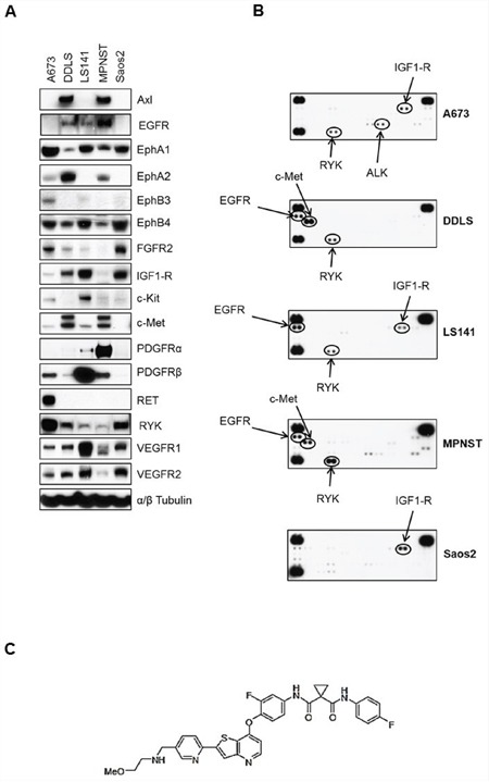
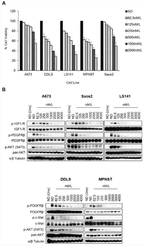
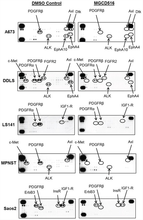
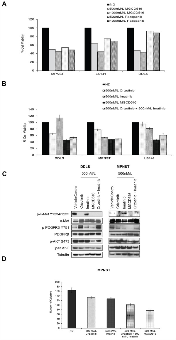
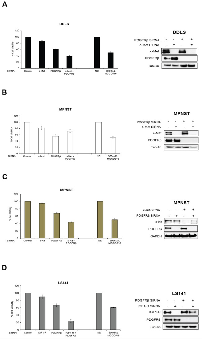
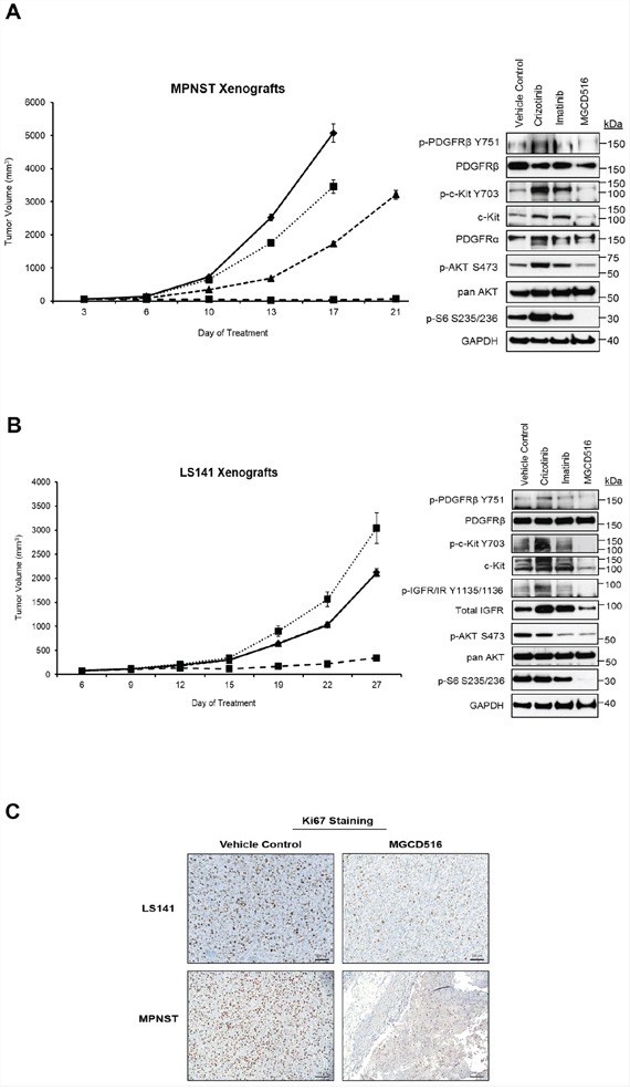
Sarcomas are rare but highly aggressive mesenchymal tumors with a median survival of 10-18 months for metastatic disease. Mutation and/or overexpression of many receptor tyrosine kinases (RTKs) including c-Met, PDGFR, c-Kit and IGF1-R drive defective signaling pathways in sarcomas. MGCD516 (Sitravatinib) is a novel small molecule inhibitor targeting multiple RTKs involved in driving sarcoma cell growth. In the present study, we evaluated the efficacy of MGCD516 both in vitro and in mouse xenograft models in vivo. MGCD516 treatment resulted in significant blockade of phosphorylation of potential driver RTKs and induced potent anti-proliferative effects in vitro. Furthermore, MGCD516 treatment of tumor xenografts in vivo resulted in significant suppression of tumor growth. Efficacy of MGCD516 was superior to imatinib and crizotinib, two other well-studied multi-kinase inhibitors with overlapping target specificities, both in vitro and in vivo. This is the first report describing MGCD516 as a potent multi-kinase inhibitor in different models of sarcoma, superior to imatinib and crizotinib. Results from this study showing blockade of multiple driver signaling pathways provides a rationale for further clinical development of MGCD516 for the treatment of patients with soft-tissue sarcoma.

Keywords: MGCD516; Sitravatinib; crizotinib; imatinib; tyrosine kinase.


Conflict of interest statement

CONFLICTS OF INTEREST

The authors have no conflicts to disclose in relation to this work.

Figures

Figure 1
Figure 1. Basal levels of total and phosphorylated receptor tyrosine kinases in five sarcoma cell lines
A. Approximately, 1 × 10⁶ cells were plated in 60 mm plates in media containing 10%FBS and grown for 24 hours. Next day, 30 micrograms of RIPA lysates were loaded on SDS/PAGE and immunoblotted using indicated antibodies. B. 1 × 10⁶ cells were plated in 60 mm plates for 24 hours in media containing 10%FBS. Next day, lysates were prepared according to manufacturer's instructions (R&D Systems, ARY001B) and 200 micrograms of lysates were applied to phospho-RTK membranes overnight at 4°C. Arrows indicate phosphorylated RTK spots in duplicate. C. MGCD516 (Sitravatinib) is a novel, potent multi-kinase small molecule inhibitor. Chemical structure of MGCD516 is shown.
Figure 2
Figure 2. MGCD516 has potent anti-proliferative effect and inhibits multiple RTKs at low nanomolar concentrations
A. Indicated cell lines were plated in 96-well plates and treated in six wells per condition with increasing doses of MGCD516 for 72 hours. Cell viability was measured using Dojindo Cell Counting Kit 8. Dose response graphs were generated as a percentage of the no drug control. Error bars represent standard error mean. Combined data from three independent experiments is shown. B. Indicated cell lines were grown to 60% confluency in serum free media overnight. Next day, cells were treated with indicated concentrations of MGCD516 in serum free media for 3 hours. After the drug treatments, cells were stimulated in drug free media containing 10%FBS. Media containing no FBS was used as unstimulated control. 20 to 30 micrograms of RIPA lysates were loaded on SDS/PAGE and immunoblotted using indicated antibodies. Representative western blots from two independent experiments are shown. ND=No Drug, Uns=Unstimulated control.
Figure 3
Figure 3. Broad spectrum activity of MGCD516 shows blockade of multiple RTKs across various sarcoma subtypes
Approximately, 1 × 10⁶ cells were plated in 60mm plates in serum free media for 24 hours. Next day, DMSO or MGCD516 was added in serum free media and treated for 3 hours. Cells were then stimulated in media containing 10% FBS for 1 hour. Following stimulation, lysates were prepared according to manufacturer's instructions (R&D Systems, ARY001B) and 200 micrograms of lysates were applied to phospho-RTK membranes overnight. Arrows indicate phosphorylated RTK spots in duplicate.
Figure 4
Figure 4. MGCD516 treatment results in superior anti-proliferative effect, better inhibition of downstream targets such as p-AKT and greater reduction in colony growth when compared to imatinib and crizotinib
A. and B. Indicated cell lines were plated in 96-well plates and treated in triplicate with indicated concentrations of drugs for 72 hours. Cell viability was measured using Dojindo Cell Counting Kit 8. Graphs were generated as a percentage of the no drug (ND) control. Error bars represent standard error mean. Combined data from at least two independent experiments is shown. Note: For Figure 4A, error bars are included; however, they are too small to be seen. C. Indicated cell lines were grown to 60% confluency in serum free media overnight. Next day, cells were treated with indicated concentrations of MGCD516 in serum free media for 3 hours. After the drug treatments, cells were stimulated in drug free media containing 10%FBS. 30 micrograms of RIPA lysates were loaded on SDS/PAGE and immunoblotted using indicated antibodies. Representative western blots from two independent experiments are shown. D. 1,000 cells were plated, in triplicate, onto 100mm dishes and treated the next day with the indicated drugs in media containing 10%FBS for 24 hours. Following treatment, cells were cultured in drug-free media for 10 to 14 days. Colonies were scored with ColCount software from Oxford Optronix (Abingdon, UK). Combined data from the experiment carried out in triplicate is shown.
Figure 5
Figure 5. siRNA mediated knockdown of potential driver RTKs results in inhibition of cell proliferation similar to MGCD516 treatment
A, B, C and D. Approximately, 5–7.5 × 10⁵ cells were plated and transfected with 50 nmol/L of indicated siRNAs (GE Dharmacon) the same day. 24 hours later, media was changed and cells were transfected again with 50 nmol/L siRNA. After another 24 hours, cells were harvested and approximately 2000 cells per well were plated in 96 well plates in triplicates. Remaining cells were lysed in RIPA buffer and lysates were used to confirm knockdown of protein expression by western immunoblotting. Cell viability was measured using Dojindo Cell Counting Kit 8. Cell viability assay carried out using MGCD516 treatment at 500 nmol/L is shown for comparison. Error bars represent standard error mean. Combined data from at least two independent experiments is shown.
Figure 6
Figure 6. MGCD516 treatment induces significant suppression of tumor growth and better inhibition of downstream targets than imatinib and crizotinib in vivo
A, B. Tumor growth of MPNST and LS141 xenografts treated with the indicated drugs is shown. For MPNST xenografts, treatment was stopped on day 17 for vehicle control and crizotinib treatment group (as the tumors were huge). Note: In the graph for LS141 xenografts, the tumor growth volume lines for vehicle control and imatinib treatment groups are overlapping and therefore not visible as two separate lines. 30 micrograms of RIPA lysates obtained using sample grinding kit (GE Healthcare) from xenograft tissues at the end of 3 week treatment were loaded on SDS/PAGE and immunoblotted using indicated antibodies. C. Xenograft tissues obtained from mice at the end of 3 week treatment with vehicle or MGCD516 were stained immunohistochemically using Ki67 antibody. Scale bar (100 μm) is shown in the lower right hand corner of each image. Representative image from at least 2 animals sacrificed at the end of drug treatment is shown.

References

    1. Forscher C, Mita M, Figlin R. Targeted therapy for sarcomas. Biologics: targets & therapy. 2014;8:91–105. - PMC - PubMed
    1. Nystrom LM, Reimer NB, Reith JD, Dang L, Zlotecki RA, Scarborough MT, Gibbs CP., Jr Multidisciplinary management of soft tissue sarcoma. TheScientificWorldJournal. 2013;2013:852462. - PMC - PubMed
    1. Clark MA, Fisher C, Judson I, Thomas JM. Soft-tissue sarcomas in adults. The New England journal of medicine. 2005;353:701–711. - PubMed
    1. Cormier JN, Pollock RE. Soft tissue sarcomas. CA. 2004;54:94–109. - PubMed
    1. Radaelli S, Stacchiotti S, Casali PG, Gronchi A. Emerging therapies for adult soft tissue sarcoma. Expert review of anticancer therapy. 2014;14:689–704. - PubMed

Publication types

MeSH terms