

Peliosis hepatis: Personal experience and literature review
- PMID: 26675327
- PMCID: PMC4674738
- DOI: 10.3748/wjg.v21.i46.13188
Peliosis hepatis: Personal experience and literature review
Abstract
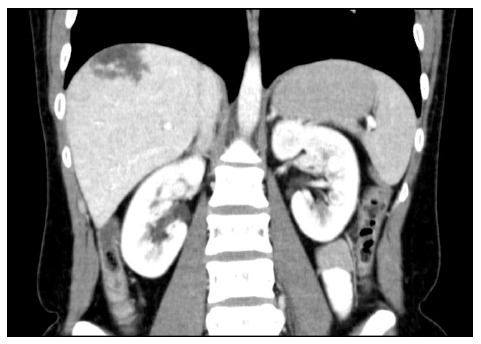
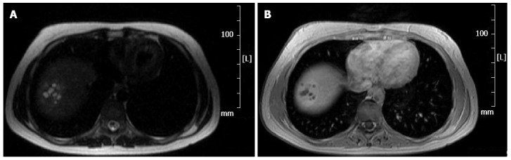
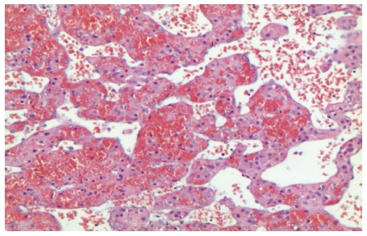
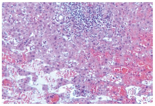
Peliosis hepatis (PH) is a disease characterized by multiple and small, blood-filled cysts within the parenchymatous organs. PH is a very rare disease, more common in adults, and when it affects the liver, it comes to the surgeon's attention only in an extremely urgent situation after the lesion's rupture with the resulting hemoperitoneum. This report describes the case of a 29-year-old woman affected by recurring abdominal pain. Computed tomography scans showed a hepatic lesion formed by multiple hypodense areas, which showed an early acquisition of the contrast during the arterial phase. Furthermore, it remained isodense with the remaining parenchyma during the late venous phase. We decided on performing a liver resection of segment VII while avoiding a biopsy for safety reasons. The histopathologic examination confirmed the diagnosis of focal PH. PH should always be considered in the differential diagnosis of hepatic lesions. Clinicians should discuss the possible causes and issues related to the differential diagnosis in addition to the appropriate therapeutic approach. The fortuitous finding of a lesion, potentially compatible with PH, requires elective surgery with diagnostic and therapeutic intents. The main aim is to prevent the risk of a sudden bleeding that, in absence of properly equipped structures, may have a fatal outcome.
Keywords: Hemoperitoneum; Hemorrhagic hepatic cysts; Liver mass; Peliosis hepatis; Surgical treatment.
Figures
References
Publication types
MeSH terms
LinkOut - more resources
Full Text Sources
Other Literature Sources
