

Retinal fibrosis in diabetic retinopathy
- PMID: 26675403
- PMCID: PMC4683353
- DOI: 10.1016/j.exer.2015.04.004
Retinal fibrosis in diabetic retinopathy
Abstract
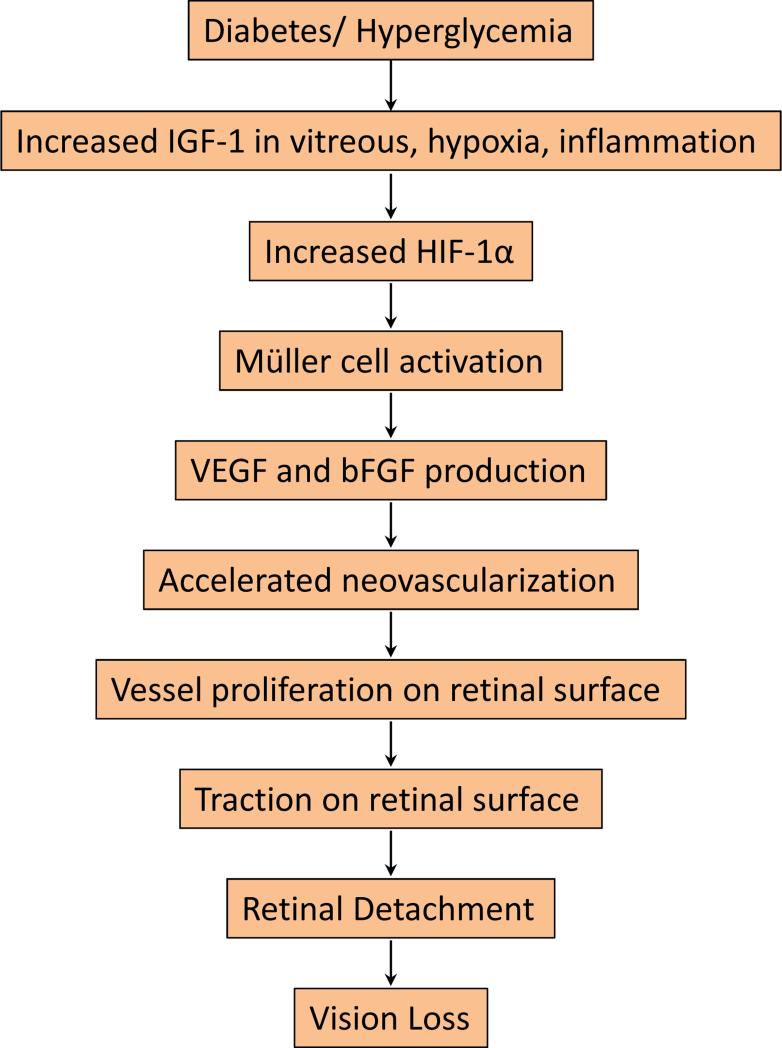
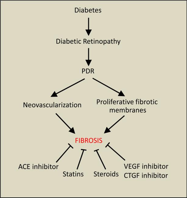
In response to injury, reparative processes are triggered to restore the damaged tissue; however, such processes are not always successful in rebuilding the original state. The formation of fibrous connective tissue is known as fibrosis, a hallmark of the reparative process. For fibrosis to be successful, delicately balanced cellular events involving cell proliferation, cell migration, and extracellular matrix (ECM) remodeling must occur in a highly orchestrated manner. While successful repair may result in a fibrous scar, this often restores structural stability and functionality to the injured tissue. However, depending on the functionality of the injured tissue, a fibrotic scar can have a devastating effect. For example, in the retina, fibrotic scarring may compromise vision and ultimately lead to blindness. In this review, we discuss some of the retinal fibrotic complications and highlight mechanisms underlying the development of retinal fibrosis in diabetic retinopathy.
Keywords: Diabetic retinopathy; Extracellular matrix; Müller cell; Retinal fibrosis; Vascular endothelial growth factor.
Copyright © 2015 Elsevier Ltd. All rights reserved.
Figures
References
-
- Alasil T, Waheed NK. Pan retinal photocoagulation for proliferative diabetic retinopathy: pattern scan laser versus argon laser. Curr Opin Ophthalmol. 2014;25(3):164–170. - PubMed
-
- Amin RH, Frank RN, Kennedy A, Eliott D, Puklin JE, Abrams GW. Vascular endothelial growth factor is present in glial cells of the retina and optic nerve of human subjects with nonproliferative diabetic retinopathy. Invest Ophthalmol Vis Sci. 1997;38(1):36–47. - PubMed
Publication types
MeSH terms
Substances
Grants and funding
LinkOut - more resources
Full Text Sources
Other Literature Sources
Medical
