. 2015 Mar 11:3:233-42.
doi: 10.1016/j.bbacli.2015.03.002. eCollection 2015 Jun.

Truncated HSPB1 causes axonal neuropathy and impairs tolerance to unfolded protein stress

Affiliations

Truncated HSPB1 causes axonal neuropathy and impairs tolerance to unfolded protein stress

Emil Ylikallio et al. BBA Clin. .

Abstract

Background: HSPB1 belongs to the family of small heat shock proteins (sHSP) that have importance in protection against unfolded protein stress, in cancer cells for escaping drug toxicity stress and in neurons for suppression of protein aggregates. sHSPs have a conserved α-crystalline domain (ACD), flanked by variable N- and C-termini, whose functions are not fully understood. Dominant missense variants in HSPB1, locating mostly to the ACD, have been linked to inherited neuropathy.

Methods: Patients underwent detailed clinical and neurophysiologic characterization. Disease causing variants were identified by exome or gene panel sequencing. Primary patient fibroblasts were used to investigate the effects of the dominant defective HSPB1 proteins.

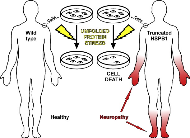
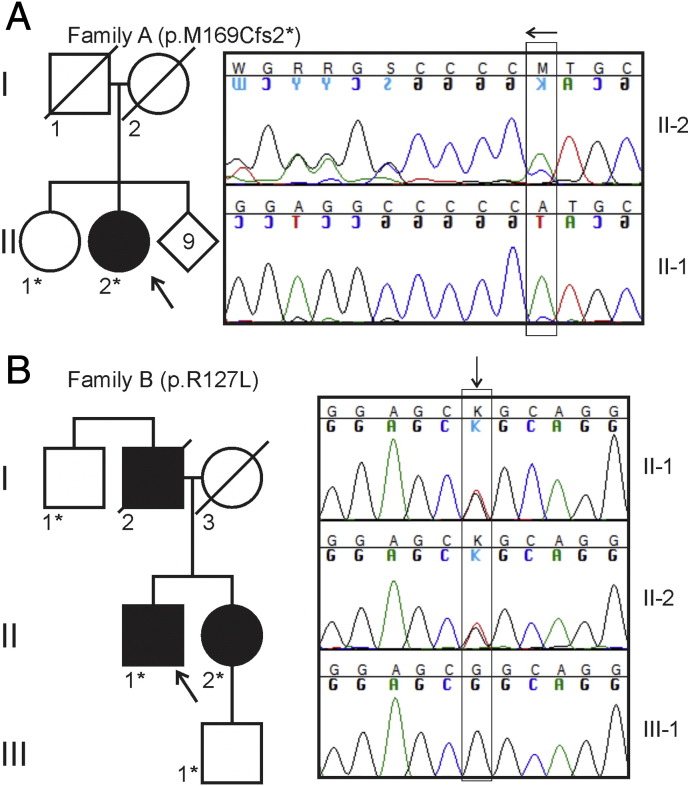
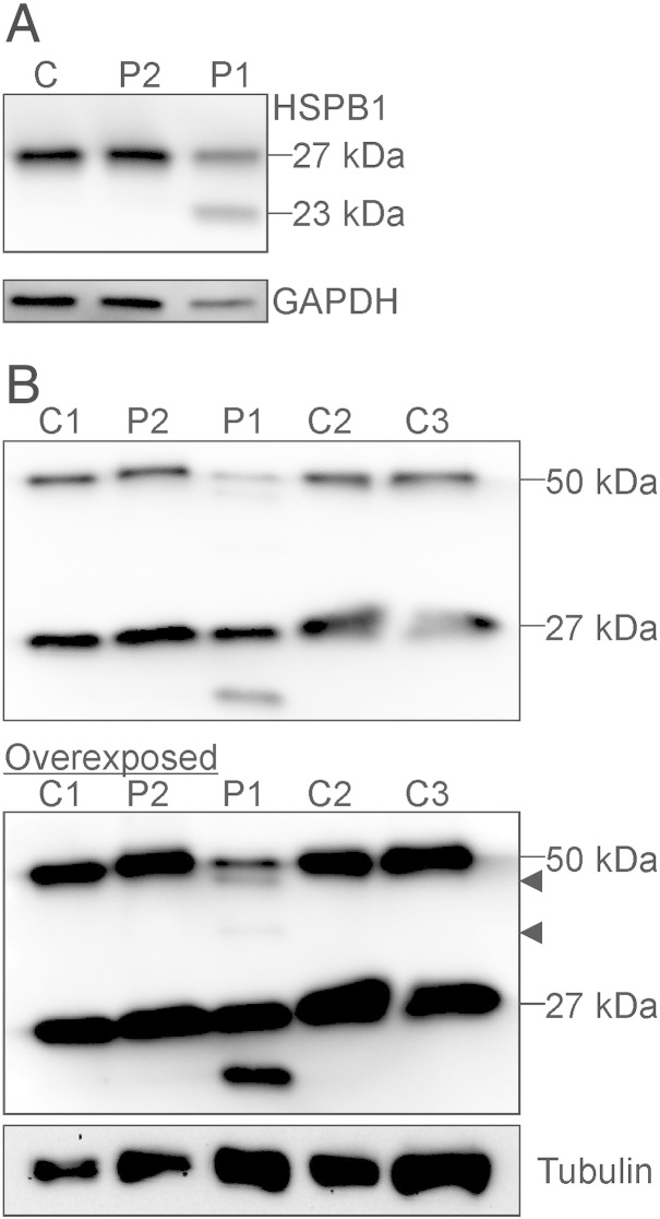
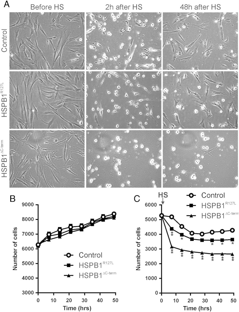
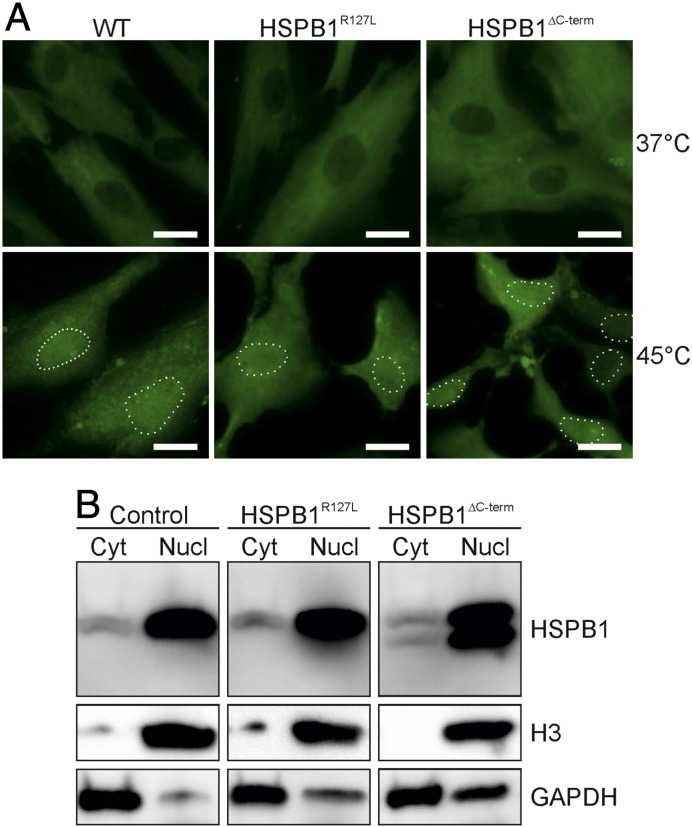
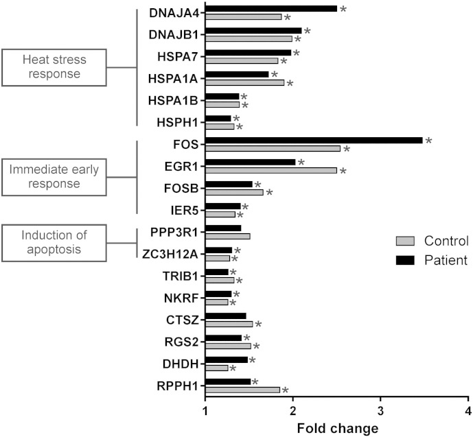
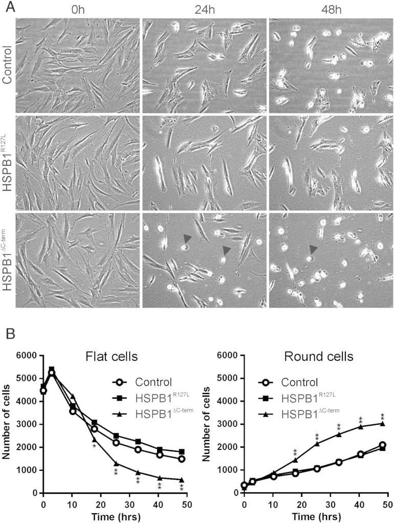
Results: Frameshift variant predicting ablation of the entire C-terminus p.(Met169Cfs2*) of HSPB1 and a missense variant p.(Arg127Leu) were identified in patients with dominantly inherited motor-predominant axonal Charcot-Marie-Tooth neuropathy. We show that the truncated protein is stable and binds wild type HSPB1. Both mutations impaired the heat stress tolerance of the fibroblasts. This effect was particularly pronounced for the cells with the truncating variant, independent of heat-induced nuclear translocation and induction of global transcriptional heat response. Furthermore, the truncated HSPB1 increased cellular sensitivity to protein misfolding.

Conclusion: Our results suggest that truncation of the non-conserved C-terminus impairs the function of HSPB1 in cellular stress response.

General significance: sHSPs have important roles in prevention of protein aggregates that induce toxicity. We showed that C-terminal part of HSPB1 is critical for tolerance of unfolded protein stress, and when lacking causes axonal neuropathy in patients.

Keywords: ACD, α-crystalline domain; CADD, combined annotation dependent depletion; CMT, Charcot–Marie–Tooth disease; Charcot–Marie–Tooth neuropathy; EMG, electromyography; ENMG, electroneuromyography; EVS, exome variant server; HSPB1; MUP, motor unit potential; Protein misfolding; QST, quantitative sensory testing; SISu, Sequencing Initiative Suomi; dHMN, distal hereditary motor neuropathy; heat shock protein; sHSP, small heat shock protein.


Figures

None
Graphical abstract
Fig. 1
Fig. 1
Pedigrees of the studied families. The DNA of the persons indicated by asterisk (*) was studied, persons crossed with diagonal lines were deceased. Representative capillary sequencing chromatograms are shown to the right. (A) In family A, the heterozygous deletion of a single adenine residue [c.505delA, HSPB1 (NM_001540.3)] is highlighted by the vertical box. The deletion leads to a frame shift causing superimposed curves in the sequence that follows (arrow, note that sequencing direction is right to left). (B) In family B, the heterozygous guanine to thymidine nucleotide change [c.380G > T, HSPB1 (NM_001540.3)] is indicated by the vertical box (arrow).
Fig. 2
Fig. 2
Stability and dimerization of the truncated protein. Lysates from primary fibroblasts were analyzed by Western blotting and immunodetection with anti-HSPB1 antibody. (A) Shown is a representative blot form control (C), HSPB1R127L (P2) and HSPB1∆C-term (P1) fibroblasts. The wild type HSPB1 protein of 27 kDa size is detected in all samples. In HSPB1∆C-term fibroblasts an additional band about 23 kDa in size, corresponding to the truncated protein, is observed. (B) Western blots under non-reducing conditions were performed to detect HSPB1 dimers. At normal exposure the control fibroblasts (C1–3) show a band at ~ 27 kDa corresponding to the wild type monomer and at ~ 50 kDa corresponding to the wild type dimer. The HSPB1R127L fibroblasts (P2) are similar to wild type. In HSPB1∆C-term fibroblasts (P1), the ~ 27 kDa monomer and ~ 50 kDa dimer can be detected, but overexposure of the same blot shows two additional fainter bands (arrowheads) corresponding to dimers formed by wild type and truncated protein or two truncated proteins.
Fig. 3
Fig. 3
Cell growth and survival after heat stress. The growth of the fibroblasts was monitored under normal culture conditions. Then the cells were subjected to 45 °C for 2 h (HS), followed by washing out of floating dead cells and automated cell monitoring for 48 h. (A) Representative images are shown for each condition in control, HSPB1R127L and HSPB1∆C-term fibroblasts. Based on cell morphology, the control and HSPB1R127L cells had mostly recovered 48 h after heat treatment. (B) Under normal conditions, cell growth of all three lines was identical. (C) After heat stress (HS, arrow) cell survival and recovery of HSPB1R127L and HSPB1∆C-term fibroblasts were worse than of control. The impairment was significantly more marked in HSPB1∆C-term fibroblasts compared to HSPB1R127L (P < 0.0001). Asterisks denotes significant differences compared to control fibroblasts *P < 0.01, **P < 0.0001 (two-way ANOVA).
Fig. 4
Fig. 4
Nuclear translocation of HSPB1. (A) Immunocytochemistry of primary fibroblasts using the anti-HSPB1 antibody (green) showed that part of the cytoplasmic protein translocates to the nucleus in 45 °C in control (WT), HSPB1∆C-term and HSPB1R127L fibroblasts. Nuclei are highlighted with dotted lines. Bars = 20 μm. (B) Nuclear enrichment was performed on cells at 45 °C, followed by Western blotting to detect HSPB1. Histone H3 protein was detected as a nuclear loading control, and GAPDH as a cytoplasmic loading control. The abundance of HSPB1 was strongly increased in the nucleus (Nucl) compared to cytoplasm (Cyt) at 45 °C. Note that the HSPB1∆C-term protein is detectable and concentrated in the nucleus following heat stress, similar to the wild type protein.
Fig. 5
Fig. 5
Gene expression changes in response to heat stress. Control and HSPB1∆C-term (patient) fibroblasts were treated by exposure to 45 °C for 30 min and a gene expression microarray was used to measure the alterations in gene expression between treated and untreated cells. Genes that were upregulated by at least 25% in both control and patient cells are shown and categorized into biological pathways. The results show that the early transcriptional response to heat was not altered in the HSPB1∆C-term fibroblasts. Asterisk indicates p-value < 0.05.
Fig. 6
Fig. 6
Sensitivity to protein misfolding. The fibroblasts were exposed to canavanine and followed by continuous cell monitoring. Representative images are shown in (A) and quantifications in (B). After 24 h of canavanine treatment, HSPB1∆C-term fibroblasts started to die (rounded cells, arrowheads) and after 48 h the same was seen for control and HSPB1R127L fibroblasts. Quantification showed that the number of cells with normal flat morphology decreased in all cell lines but significantly more in HSPB1∆C-term fibroblasts. Conversely, the number of dying (round) cells increased significantly more in HSPB1∆C-term fibroblasts compared to the other cell lines. Asterisks denotes significant difference compared to control fibroblasts *P < 0.01, **P < 0.0001 (two-way ANOVA).

References

    1. Szigeti K., Lupski J.R. Charcot–Marie–Tooth disease. Eur. J. Hum. Genet. 2009;17:703–710. - PMC - PubMed
    1. Rossor A.M., Kalmar B., Greensmith L., Reilly M.M. The distal hereditary motor neuropathies. J. Neurol. Neurosurg. Psychiatry. 2012;83:6–14. - PubMed
    1. Rossor A.M., Polke J.M., Houlden H., Reilly M.M. Clinical implications of genetic advances in Charcot–Marie–Tooth disease. Nat. Rev. Neurol. 2013;9:562–571. - PubMed
    1. Mymrikov E.V., Seit-Nebi A.S., Gusev N.B. Large potentials of small heat shock proteins. Physiol. Rev. 2011;91:1123–1159. - PubMed
    1. Irobi J., Van Impe K., Seeman P., Jordanova A., Dierick I., Verpoorten N., Michalik A., De Vriendt E., Jacobs A., Van Gerwen V., Vennekens K., Mazanec R., Tournev I., Hilton-Jones D., Talbot K., Kremensky I., Van Den Bosch L., Robberecht W., Van Vandekerckhove J., Van Broeckhoven C., Gettemans J., De Jonghe P., Timmerman V. Hot-spot residue in small heat-shock protein 22 causes distal motor neuropathy. Nat. Genet. 2004;36:597–601. - PubMed