

Overview of left ventricular outpouchings on cardiac magnetic resonance imaging
- PMID: 26675616
- PMCID: PMC4666692
- DOI: 10.3978/j.issn.2223-3652.2015.11.02
Overview of left ventricular outpouchings on cardiac magnetic resonance imaging
Abstract
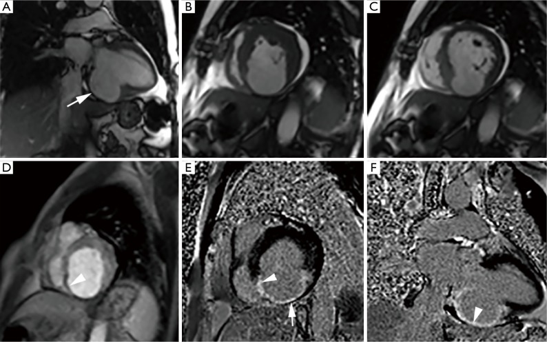
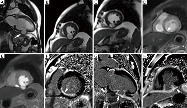
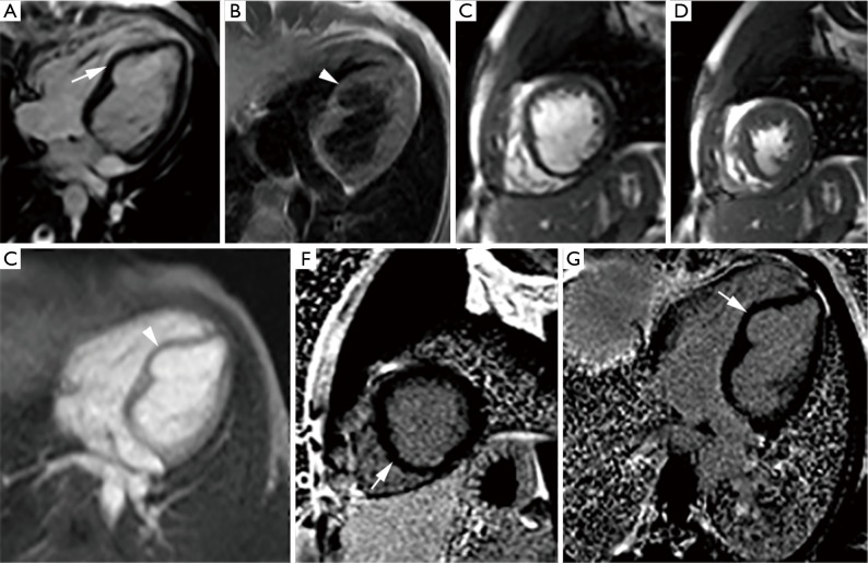
Left ventricular outpouchings commonly include aneurysm, pseudoaneurysm, and diverticulum and are now being increasingly detected on imaging. Distinction between these entities is of prime importance to guide proper management as outcomes for these entities differ substantially. Chest radiograph is usually nonspecific in their diagnosis. Echocardiography, multi-detector computed tomography evaluation and angiography are helpful in the diagnosis with their inherit limitations. Cardiac magnetic resonance imaging (MRI) is emerging as a very useful tool that allows simultaneous anatomical and functional evaluation along with tissue characterization, which has diagnostic, theraputic and prognostic implications. This article gives an overview of left ventricular outpouchings with special emphasis on their differentiation using cardiac MRI.
Keywords: Diverticulum; aneurysm; magnetic resonance imaging (MRI); pseudoaneurysm.
Conflict of interest statement
Figures
References
-
- Heatlie GJ, Mohiaddin R. Left ventricular aneurysm: comprehensive assessment of morphology, structure and thrombus using cardiovascular magnetic resonance. Clin Radiol 2005;60:687-92. - PubMed
-
- Cho MN, Mehta SK, Matulevicius S, et al. Differentiating true versus pseudo left ventricular aneurysm: a case report and review of diagnostic strategies. Cardiol Rev 2006;14:e27-30. - PubMed
-
- Frances CD, Shlipak MG, Grady D. Left ventricular pseudoaneurysm: diagnosis by cine magnetic resonance imaging. Cardiology 1999;92:217-9. - PubMed
-
- Uribe S, Muthurangu V, Boubertakh R, et al. Whole-heart cine MRI using real-time respiratory self-gating. Magn Reson Med 2007;57:606-13. - PubMed
-
- Buehrer M, Curcic J, Boesiger P, et al. Prospective self-gating for simultaneous compensation of cardiac and respiratory motion. Magn Reson Med 2008;60:683-90. - PubMed
Publication types
LinkOut - more resources
Full Text Sources