. 2016 Jan 14;529(7585):221-5.
doi: 10.1038/nature16161. Epub 2015 Dec 14.

Tuft-cell-derived IL-25 regulates an intestinal ILC2-epithelial response circuit

Affiliations

Tuft-cell-derived IL-25 regulates an intestinal ILC2-epithelial response circuit

Jakob von Moltke et al. Nature. .

Abstract

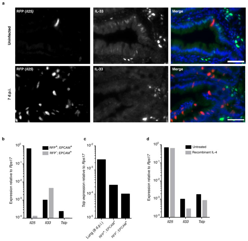
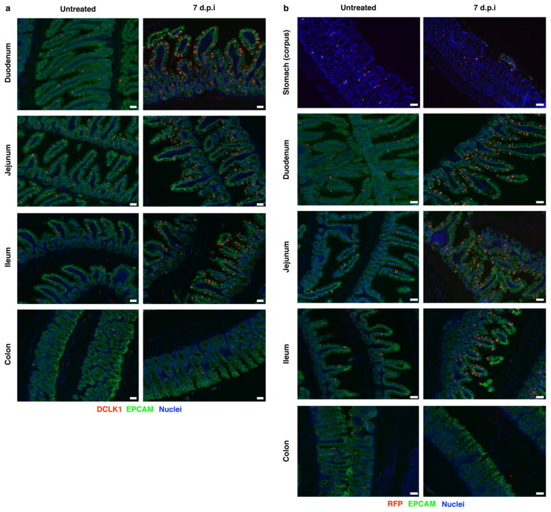
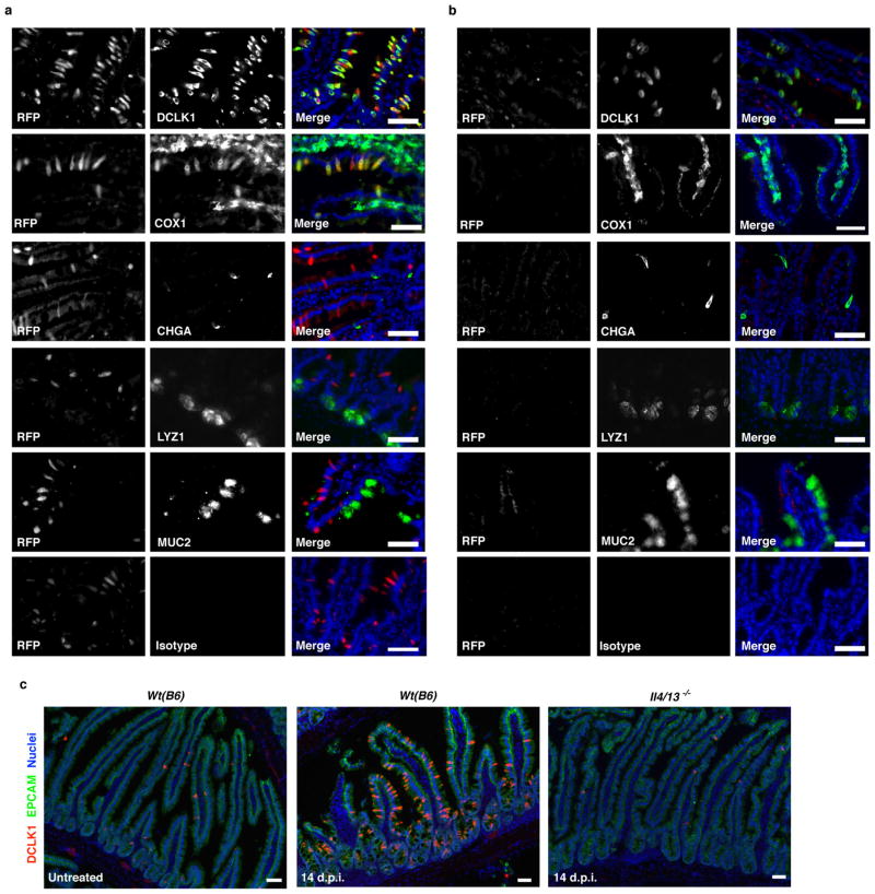
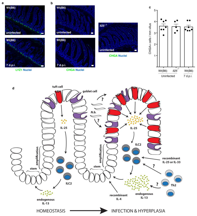
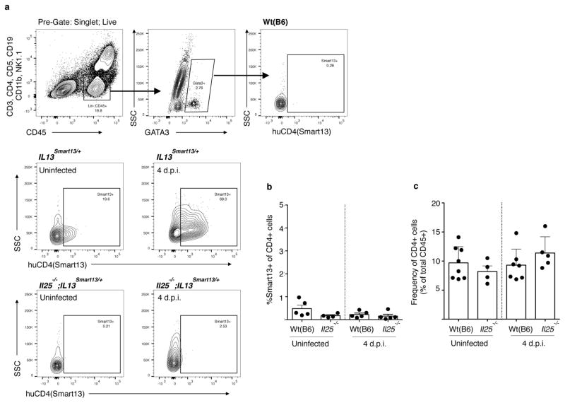
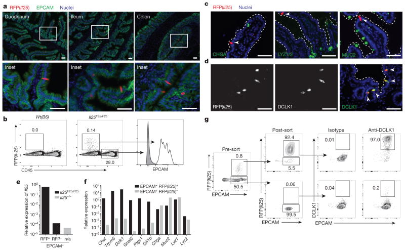
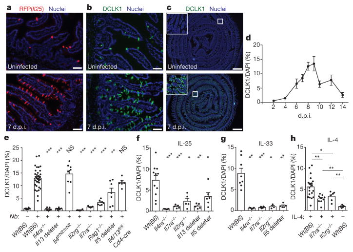
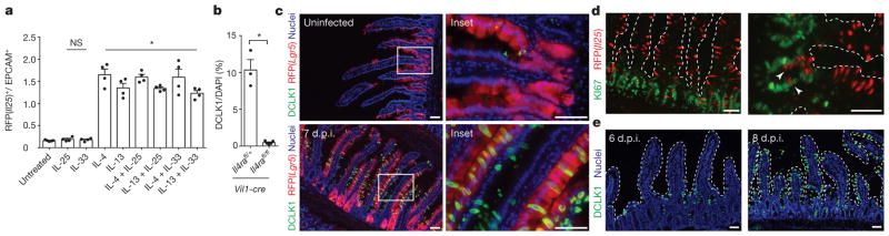
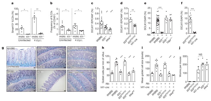
Parasitic helminths and allergens induce a type 2 immune response leading to profound changes in tissue physiology, including hyperplasia of mucus-secreting goblet cells and smooth muscle hypercontractility. This response, known as 'weep and sweep', requires interleukin (IL)-13 production by tissue-resident group 2 innate lymphoid cells (ILC2s) and recruited type 2 helper T cells (TH2 cells). Experiments in mice and humans have demonstrated requirements for the epithelial cytokines IL-33, thymic stromal lymphopoietin (TSLP) and IL-25 in the activation of ILC2s, but the sources and regulation of these signals remain poorly defined. In the small intestine, the epithelium consists of at least five distinct cellular lineages, including the tuft cell, whose function is unclear. Here we show that tuft cells constitutively express IL-25 to sustain ILC2 homeostasis in the resting lamina propria in mice. After helminth infection, tuft-cell-derived IL-25 further activates ILC2s to secrete IL-13, which acts on epithelial crypt progenitors to promote differentiation of tuft and goblet cells, leading to increased frequencies of both. Tuft cells, ILC2s and epithelial progenitors therefore comprise a response circuit that mediates epithelial remodelling associated with type 2 immunity in the small intestine, and perhaps at other mucosal barriers populated by these cells.


Conflict of interest statement

The authors declare no competing financial interests.

Figures

Extended Data Figure 1
Extended Data Figure 1. Flare25 mouse and Vil1-cre-mediated Il25 deletion
a, Gene-targeting strategy for the flox and reporter of Il25 (Flare25) mouse. b, PCR of genomic DNA isolated from the tail (lane 1, 2) or cells sorted from the small intestine (lane 3, 4) of indicated mice. c, Quantitative RT–PCR for Il25 on cDNA from EPCAM+ cells sorted from the small intestine of indicated mice. b, c, Data are representative of two experiments (n = 2). Frt, target site for FLIPASE recombinase; IRES, internal ribosomal entry site; loxP, target site for Cre recombinase; pA, bovine growth hormone poly(A) tail; tdRFP,3tandem-dimer red fluorescent protein; UTR, untranslated region. For gel source data (b) see Supplementary Fig. 1.
Extended Data Figure 2
Extended Data Figure 2. Il25 expression in epithelial surfaces
a, b, Indicated tissues of Il25F25/F25 (a) and wild-type control (b) mice stained by immunohistochemistry for RFP (red), EPCAM (green), and DAPI (blue). Some data from Fig. 1a are repeated here to allow complete comparison. Scale bars, 50 μm. Images are representative of at least three independent experiments. n =3.
Extended Data Figure 3
Extended Data Figure 3. Flow cytometry gating strategies and organoid culture
a, Flow cytometric analysis of indicated tissues in Il25F25/F25 and wild-type mice. b, Flow cytometric analysis of small intestine epithelial cells of Il25F25/F25 mice before and after fluorescence-activated cell sorting (FACS) into RFP+EPCAM+ and RFPEPCAM+ pools for analysis by quantitative RT–PCR. ce, Representative flow cytometric analysis of small-intestine-derived organoids from Il25F25/F25 (ce) and wild-type (c) mice cultured with or without recombinant protein (20 ng ml−1), as indicated (ce) or Notch signalling inhibitor DAPT (25 μM) (e). Single-cell suspensions of the organoids were stained for EPCAM (ce) and DCLK1 (d), and gated to quantify tuft cell (RFP+EPCAM+ or DCLK1+EPCAM+) frequency. f, Quantification of two technical replicates from experiment shown in e. d.p.i., days post-N. brasiliensis infection. Data in f are technical replicates. Data are representative of three (a, b, d) or two (c, e, f) independent experiments. In ad, n =3; in e, f, n =2. Error bars represent mean ±s.e.m.
Extended Data Figure 4
Extended Data Figure 4. Il25 is expressed constitutively in tuft cells
ac, Jejunum (a, b) or trachea (c) of Il25F25/F25 (a, c) and wild-type control (b) mice stained by immunohistochemistry for RFP (red), indicated lineage markers (green), and DAPI (blue). Scale bars, 50 μm. Images are representative of one (c) or two (a, b) independent experiments. n =2.
Extended Data Figure 5
Extended Data Figure 5. Tuft cells are not a major source of intestinal TSLP or IL-33
a, Jejunum of Il25F25/F25 mice stained for RFP (red), IL-33 (green), and DAPI (blue). bd, Quantitative RT–PCR on indicated (b, c) or RFP+EPCAM+ (d) cells sorted from untreated (b, c) mice or mice treated as indicated (d). RNA isolated from whole lung 8 days post-N. brasiliensis infection is used as a positive control for Tslp expression in c. Expression of Tslp in sorted Tslp-expressing cells of the lung would probably be higher. Scale bars, 50 μm. Data are representative of two independent experiments. In a, n =3; in bd, n =2.
Extended Data Figure 6
Extended Data Figure 6. N. brasiliensis induces tuft cell hyperplasia throughout the small intestine but not in stomach and colon
a, b, Indicated tissues of Il25F25/F25 (a) and wild-type control (b) mice treated as indicated and stained by immunohistochemistry for RFP (a) or DCLK1 (b) (red), EPCAM (green), and DAPI (blue). d.p.i., days post-N. brasiliensis infection. Scale bars, 50 μm. Data are representative of two (stomach and colon) or at least three (all others) independent experiments. In a, stomach and colon: n = 2; all others: n >5.
Extended Data Figure 7
Extended Data Figure 7. Il25 is expressed only in tuft cells during worm infection and H. polygyrus infection also induces tuft cell hyperplasia
a, b, Jejunum of Il25F25/F25 (a) and wild-type control (b) mice infected for 7 days with N. brasiliensis stained by immunohistochemistry for RFP (red), indicated lineage markers (green), and DAPI (blue). c, Jejunum of indicated mice left untreated or infected 14 days with H. polygyrus and stained by immunohistochemistry for DAPI (blue), EPCAM (green) and DCLK1 (red). Scale bars, 50 μm. d.p.i., days post-H. polygyrus infection. Images are representative of one (c) or two (a, b) independent experiments. In a, b, n =2; in c, n = 1 (uninfected) or n =2 (infected).
Extended Data Figure 8
Extended Data Figure 8. Absence of Paneth and CHGA+ cell hyperplasia after N. brasiliensis infection and model of ILC2–epithelial signalling circuit
a, b, Jejunum of indicated mice stained for DAPI (blue) and LYZ1/2 (a) or CHGA (b) (green). c, Quantification of CHGA+ cells from imaging in (b). d, During homeostasis, rare epithelial tuft cells of the small intestine constitutively express Il25, which maintains low levels of IL-13 production in lamina propria ILC2s. IL-13 in turn signals uncommitted epithelial progenitors to promote emergence of tuft and goblet cells. In the absence of infection, this feed-forward ILC2–epithelial circuit is restrained by as yet unknown mechanisms. After N. brasiliensis (N.b.) infection, a helminth-derived signal or a change in host physiology activates the ILC2–epithelial circuit leading to tuft and goblet cell hyperplasia and enhanced IL-13 production by ILC2s. Adaptive TH2 cells probably also provide IL-13 and/or support ILC2 activation, especially when infection or inflammation lasts more than a week. Recombinant proteins are sufficient to induce tuft cell hyperplasia, either by inducing IL-13 production in lymphoid cells (IL-25 or IL-33) or by directly binding epithelial progenitors (IL-4). Scale bars, 50 μ m. d.p.i., days post-N. brasiliensis infection. Data in c are biological replicates. Data are representative of two (a) or three (b) independent experiments or pooled from multiple experiments (c). In a, n =2; in b, c, n is as shown in c. Error bars represent mean ±s.e.m.
Extended Data Figure 9
Extended Data Figure 9. IL-13 production by lamina propria ILC2 and CD4+ cells
a, b, Lamina propria cells from Il25−/−;Il13Smart/+, Il13Smart/+, and wild-type control mice analysed by flow cytometry and gated on ILC2 (a, LinCD45+GATA3+) or CD45+CD4+ (b) cells. IL-13 secretion was quantified by measuring surface expression of human CD4, which is expressed from the Il13 locus in Il13Smart reporter mice. c, Frequency of lamina propria CD4+ cells as a percentage of total CD45+ cells as assessed by flow cytometry. d.p.i., days post-N. brasiliensis infection. Data in b, c are biological replicates. Data are representative of at least three (a) independent experiments, or pooled from multiple experiments (b, c). In a, n =5; in b, c, n is as shown. Error bars represent mean ±s.e.m.
Figure 1
Figure 1. Intestinal tuft cells constitutively express Il25
a, Indicated tissues from Il25F25/F25 mice stained for RFP (red), EPCAM (green) and 4′,6-diamidino-2-phenylindole (DAPI; blue). b, Flow cytometry of digested jejunum. c, d, Jejunum from Il25F25/F25 mice stained as indicated. Dotted lines outline villi. Arrowheads indicate RFP+ cells. e, f, Quantitative polymerase chain reaction with reverse transcription (RT–PCR) on cells sorted from small intestines of Il25−/− (e) and Il25F25/F25 (e, f) mice. n/a, not applicable. g, Flow cytometry of cells sorted from small intestines of Il25F25/F25 mice and stained with anti-DCLK1. Scale bars,350 μm. All data are biological replicates. Data are representative of two (bd, g), or at least three (a, e, f) experiments. In a, n >5; in bd, g, n =2; in e, f, n =3.
Figure 2
Figure 2. Worm infection induces IL-13-dependent tuft cell hyperplasia
ac, Jejunum from Il25F25/F25 mice stained for RFP (a) or DCLK1 (b, c). dh, Immunohistochemical quantification of tuft cells (DCLK1+) in duodenum/jejunum of mice infected with N. brasiliensis for indicated days (d) or 7 days (e) or injected with indicated protein (fh). Scale bars: 50 μm (a, b), 1 mm (c). All data are biological replicates. Data are representative of at least three (ac) experiments or pooled (dh) from multiple experiments. In ac, n >10; in d, day 2: n = 2; days 4, 12, 14: n = 4; day 9: n = 5; days 6, 8, 10: n = 6; day 7: n =8; in eh, n is as shown. Nb, N. brasiliensis. *P <0.05; **P <0.01, ***P < 0.001; NS, not significant (Mann–Whitney test). Error bars represent mean ±standard error of the mean (s.e.m.).
Figure 3
Figure 3. IL-13 signalling in epithelial progenitors gives rise to tuft cell hyperplasia
a, Flow cytometric quantification of RFP+ tuft cells in intestinal organoids grown from Il25F25/F25 mice and treated with indicated recombinant proteins (20 ng ml−1). b, Immunohistochemical quantification of tuft cells (DCLK1+) in duodenum/jejunum of mice infected 7 days with N. brasiliensis. c, Jejunum of Lgr5cre-Ert2/+; Gt(ROSA)26STOP-flox::RFP/+ mice treated 5 days with tamoxifen and stained for DCLK1 (green) and DAPI (blue). N. brasiliensis infection as indicated. d, e, Jejunum of Il25F25/F25 (d) or Wt(B6) (e) mice infected for 7 days (d) or indicated number of days (e) with N. brasiliensis and stained for RFP (red) and Ki67 (green) (d) or DCLK1 (green) and DAPI (blue) (e). d, e, Dotted lines outline villi. Scale bars, 50 μm. Arrowheads indicate Ki67 and RFP overlap. Data in a are technical replicates, all other data are biological replicates. Data are representative of two (ce) or three (a) experiments or pooled (b) from multiple experiments. In a, b, n is as shown; in c, uninfected: n =4; 7 d.p.i.: n =5; in d, n =2; in e, n =4. Nb, N. brasiliensis. *P < 0.05; NS, not significant (Mann–Whitney test). Error bars represent mean ±s.e.m.
Figure 4
Figure 4. Tuft cells regulate intestinal physiology through an ILC2–epithelium response circuit
a, b, Flow cytometric analysis of lamina propria ILC2s (Lin, CD45+, GATA3+) from Il13Smart/+ and Il25−/−;Il13Smart/+ mice. c, d, Flow cytometric quantification of tuft cells (DCLK1+) in jejunum of uninfected mice. e, f, Immunohistochemical quantification of tuft cells (DCLK1+) in jejunum/duodenum of mice infected 7 days with N. brasiliensis. In e, wild-type controls are the same as in Fig. 2e. g, Jejunum of mice treated as indicated and stained for goblet cells with periodic acid Schiff (PAS) Alcian blue. h, i, Goblet cell number (h) and size (i) calculated from imaging. j, Intestinal worm burden in mice infected 10 days with N. brasiliensis. Scale bars, 50 μm. All data are biological replicates. Data are representative of two (g) experiments or pooled (af, hj) from multiple experiments. In g, n is as shown in h and i; in af, hj, n is as shown. Nb, N. brasiliensis. *P <0.05; **P <0.01; ***P < 0.001; NS, not significant (Mann–Whitney test). Error bars represent mean ±s.e.m.

Comment in

References

    1. Miller HR, Nawa Y. Immune regulation of intestinal goblet cell differentiation. Specific induction of nonspecific protection against helminths? Nouv Rev Fr Hematol. 1979;21:31–45. - PubMed
    1. Castro GA, Badial-Aceves F, Smith JW, Dudrick SJ, Weisbrodt NW. Altered small bowel propulsion associated with parasitism. Gastroenterology. 1976;71:620–625. - PubMed
    1. Grencis RK. Immunity to helminths: resistance, regulation, and susceptibility to gastrointestinal nematodes. Annu Rev Immunol. 2015;33:201–225. - PubMed
    1. Price AE, et al. Systemically dispersed innate IL-13-expressing cells in type 2 immunity. Proc Natl Acad Sci USA. 2010;107:11489–11494. - PMC - PubMed
    1. Neill DR, et al. Nuocytes represent a new innate effector leukocyte that mediates type-2 immunity. Nature. 2010;464:1367–1370. - PMC - PubMed

Publication types

MeSH terms