Review
. 2016 Jan;96(1):253-305.
doi: 10.1152/physrev.00007.2015.

Absence of Dystrophin Disrupts Skeletal Muscle Signaling: Roles of Ca2+, Reactive Oxygen Species, and Nitric Oxide in the Development of Muscular Dystrophy

Affiliations
Review

Absence of Dystrophin Disrupts Skeletal Muscle Signaling: Roles of Ca2+, Reactive Oxygen Species, and Nitric Oxide in the Development of Muscular Dystrophy

David G Allen et al. Physiol Rev. 2016 Jan.

Abstract

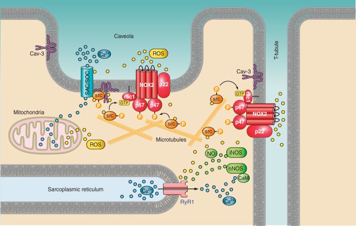
Dystrophin is a long rod-shaped protein that connects the subsarcolemmal cytoskeleton to a complex of proteins in the surface membrane (dystrophin protein complex, DPC), with further connections via laminin to other extracellular matrix proteins. Initially considered a structural complex that protected the sarcolemma from mechanical damage, the DPC is now known to serve as a scaffold for numerous signaling proteins. Absence or reduced expression of dystrophin or many of the DPC components cause the muscular dystrophies, a group of inherited diseases in which repeated bouts of muscle damage lead to atrophy and fibrosis, and eventually muscle degeneration. The normal function of dystrophin is poorly defined. In its absence a complex series of changes occur with multiple muscle proteins showing reduced or increased expression or being modified in various ways. In this review, we will consider the various proteins whose expression and function is changed in muscular dystrophies, focusing on Ca(2+)-permeable channels, nitric oxide synthase, NADPH oxidase, and caveolins. Excessive Ca(2+) entry, increased membrane permeability, disordered caveolar function, and increased levels of reactive oxygen species are early changes in the disease, and the hypotheses for these phenomena will be critically considered. The aim of the review is to define the early damage pathways in muscular dystrophy which might be appropriate targets for therapy designed to minimize the muscle degeneration and slow the progression of the disease.


Figures

FIGURE 1.
FIGURE 1.
The dystrophin protein complex (DPC). Shown are the interactions between core components of the DPC, the extracellular matrix, and nNOS. Numbers in dystrophin indicate hinge regions (H1, H2, etc.) and spectrin-like repeat domains (4, 8, 12, etc). nNOS, neuronal nitric oxide synthase; Syn, syntrophin; SSPN, sarcospan; ABD, actin binding domain; DBD, dystroglycan binding domain; SBS, syntrophin binding site; CC, coiled-coil domain; N, amino terminus; C, carboxy terminus.
FIGURE 2.
FIGURE 2.
The dystrophin molecule. Alignment of dystrophin protein structure with gene exons. Top: dystrophin protein domains with binding sites for structural and signaling proteins and lipids. ABD, actin binding domain; DBD, β-dystroglycan binding domain; SBS, syntrophin binding domain; CC, coiled-coiled region that binds α-dystrobrevin. Numbers identify spectrin-like repeats (for clarity, only 4, 8, 12, 16, 20, and 24 are indicated). Par-1b, polarity regulating kinase; AnkB, ankyrin B binding site. Bottom: corresponding exons in the human dystrophin gene. Note that the exons are arranged to correspond with the dystrophin protein structure. Therefore, the widths of the boxes do not correspond to the relative sizes of the exons. Data used to construct this figure are from the Leiden Muscular Dystrophy Pages (http://www.dmd.nl/).
FIGURE 3.
FIGURE 3.
The mechanism of stretch-induced membrane permeability in dystrophic muscle. A: according to the original “membrane tear” theory, the increased dye entry into dystrophic muscle fibers was attributed to micro-tears in the sarcolemma, caused by eccentric contractions. The membrane tear theory predicts that extracellular dyes would rapidly enter the muscle fiber immediately after the mechanically induced tear was produced, and within seconds the membrane would reseal and trap the dye inside the fiber. Therefore, the amount of dye inside the fiber would remain constant at all times after the eccentric contractions. However, experimental results indicate a different mechanism of dye entry. Extracellular dye uptake is very small immediately after the eccentric contractions and progressively increases up to at least 60 min post eccentric contractions. Moreover, both mechanosensitive channel blockers (streptomycin and Gsmtx-4) and the antioxidant N-acetyl cysteine can prevent almost all of the dye uptake, suggesting pathways involving Ca2+ entry through mechanosensitive channels and ROS produced by NOX2 are responsible for the increased membrane permeability (see text for details). B: experimental results showing the time course of procion orange dye uptake after eccentric contractions in mdx EDL muscle and the inhibitory effect of the MSC blocker streptomycin. [From Whitehead et al. (505).] C: experimental results showing the inhibitory effect of the antioxidant NAC on Evans Blue dye entry in mdx EDL muscle following eccentric contractions caused by downhill treadmill running. [From Whitehead et al. (504).]
FIGURE 4.
FIGURE 4.
Nitric oxide synthase (NOS). A: Different isoforms of NOS are generated by 3 genes. All have three domains in common: the oxygenase domain, the reductase domain, and the calcium/calmodulin binding domain (CaM). The muscle-specific form of nNOS contains an insert (μ) that is absent from the brain isoform, nNOSα. The PDZ domain in the NH2-terminal region of nNOSμ binds numerous signaling proteins. The nNOSβ NH2-terminal domain likely targets this isoform to the Golgi elements. eNOS is cotranslationally myristoylated (Myr) on the NH2 terminus and posttranslationally by palmitoylation (Palm) at two nearby sites. B: All isoforms are active as homodimers formed by interaction of the oxygenase domains. Arg, arginine binding site; BH4, tetrahydrobiopterin binding site; FMN, flavin mononucleotide binding site; FAD, flavin adenine dinucleotide binding site; NADPH, binding site for reduced form of nicotinamide adenine dinucleotide phosphate. [The figure of the NOS dimer (B) was kindly provided by Dr. Philip Dash, Univ. of Reading, UK.]
FIGURE 5.
FIGURE 5.
Mechanisms of stretch-induced reactive oxygen species (ROS) production and Ca2+ influx in dystrophic muscle. This schematic highlights some of the key deleterious pathways that lead to impaired muscle function, resulting from the loss of dystrophin. Particular focus is given to the pathways relating to ROS, Ca2+ and NO. During eccentric contractions, NADPH oxidase 2 (NOX2) is stimulated by a pathway involving microtubules and activation of rac1 and src kinase (src). This leads to phosphorylation of Nox subunits and activation of Nox2. The ROS produced by Nox2 further activates src, which triggers the opening of stretch-activated or store-operated channels (SAC/SOC), and influx of Ca2+. Increased Ca2+ uptake by mitochondria can also stimulate additional ROS production. Both ROS and NO, produced by mislocalized nNOS and/or iNOS, increase the opening of the RyR, leading to Ca2+ leak. See text for further details.
FIGURE 6.
FIGURE 6.
Mechanisms of damage and functional impairment in dystrophic muscle. This schematic highlights some of the key deleterious pathways that lead to impaired muscle function, as a result of the loss of dystrophin. Particular focus is given to the pathways relating to ROS, Ca2+, NO, and fibrosis. See text for details.
FIGURE 7.
FIGURE 7.
Therapeutic pathways for Duchenne muscular dystrophy (DMD). This figure highlights some of the key therapeutic targets for DMD, which include both muscle-specific and extracellular pathways. A wide range of therapeutic approaches are being evaluated for DMD (red boxes). These include gene-based therapies, pharmacological agents, and recombinant proteins (see text for details).

Similar articles

Cited by

References

    1. Aartsma-Rus A, Muntoni F. 194th ENMC International Workshop. 3rd ENMC workshop on exon skipping: towards clinical application of antisense-mediated exon skipping for Duchenne muscular dystrophy 8–10 December 2012, Naarden, The Netherlands. Neuromuscul Disord 23: 934–944, 2013. - PubMed
    1. Abramovici H, Gee SH. Morphological changes and spatial regulation of diacylglycerol kinase-zeta, syntrophins, and Rac1 during myoblast fusion. Cell Motil Cytoskeleton 64: 549–567, 2007. - PubMed
    1. Abramovici H, Hogan AB, Obagi C, Topham MK, Gee SH. Diacylglycerol kinase-zeta localization in skeletal muscle is regulated by phosphorylation and interaction with syntrophins. Mol Biol Cell 14: 4499–4511, 2003. - PMC - PubMed
    1. Adamo CM, Dai DF, Percival JM, Minami E, Willis MS, Patrucco E, Froehner SC, Beavo JA. Sildenafil reverses cardiac dysfunction in the mdx mouse model of Duchenne muscular dystrophy. Proc Natl Acad Sci USA 107: 19079–19083, 2010. - PMC - PubMed
    1. Adams ME, Butler MH, Dwyer TM, Peters MF, Murnane AA, Froehner SC. Two forms of mouse syntrophin, a 58 kd dystrophin-associated protein, differ in primary structure and tissue distribution. Neuron 11: 531–540, 1993. - PubMed

Publication types

MeSH terms