. 2016 Jan;5(1):8-19.
doi: 10.5966/sctm.2015-0157. Epub 2015 Dec 16.

Naïve Induced Pluripotent Stem Cells Generated From β-Thalassemia Fibroblasts Allow Efficient Gene Correction With CRISPR/Cas9

Affiliations

Naïve Induced Pluripotent Stem Cells Generated From β-Thalassemia Fibroblasts Allow Efficient Gene Correction With CRISPR/Cas9

Yuanyuan Yang et al. Stem Cells Transl Med. 2016 Jan.

Erratum in

Abstract

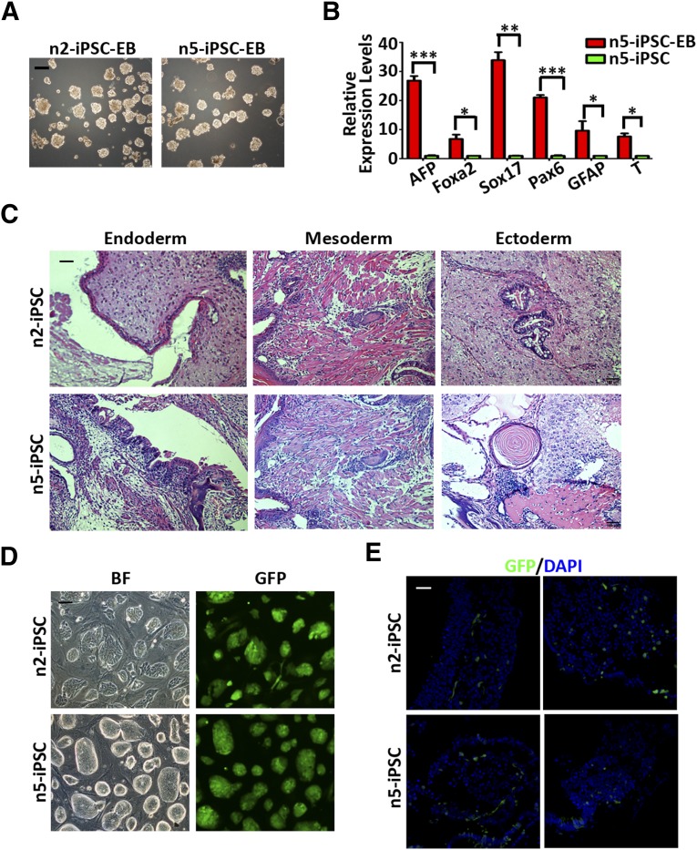
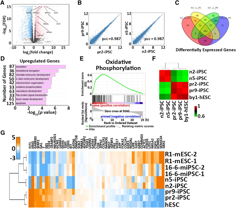
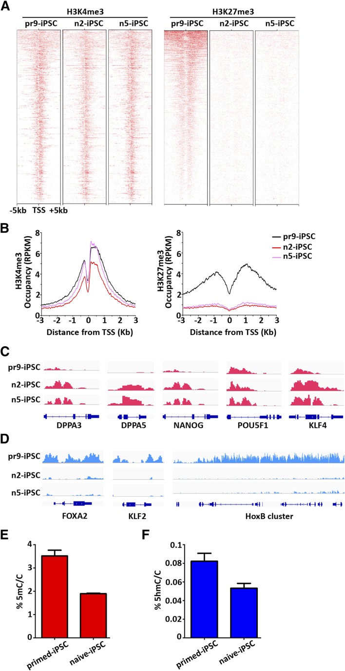
Conventional primed human embryonic stem cells and induced pluripotent stem cells (iPSCs) exhibit molecular and biological characteristics distinct from pluripotent stem cells in the naïve state. Although naïve pluripotent stem cells show much higher levels of self-renewal ability and multidifferentiation capacity, it is unknown whether naïve iPSCs can be generated directly from patient somatic cells and will be superior to primed iPSCs. In the present study, we used an established 5i/L/FA system to directly reprogram fibroblasts of a patient with β-thalassemia into transgene-free naïve iPSCs with molecular signatures of ground-state pluripotency. Furthermore, these naïve iPSCs can efficiently produce cross-species chimeras. Importantly, using the clustered regularly interspaced short palindromic repeats (CRISPR)/CRISPR-associated protein 9 nuclease genome editing system, these naïve iPSCs exhibit significantly improved gene-correction efficiencies compared with the corresponding primed iPSCs. Furthermore, human naïve iPSCs could be directly generated from noninvasively collected urinary cells, which are easily acquired and thus represent an excellent cell resource for further clinical trials. Therefore, our findings demonstrate the feasibility and superiority of using patient-specific iPSCs in the naïve state for disease modeling, gene editing, and future clinical therapy.

Significance: In the present study, transgene-free naïve induced pluripotent stem cells (iPSCs) directly converted from the fibroblasts of a patient with β-thalassemia in a defined culture system were generated. These naïve iPSCs, which show ground-state pluripotency, exhibited significantly improved single-cell cloning ability, recovery capacity, and gene-targeting efficiency compared with conventional primed iPSCs. These results provide an improved strategy for personalized treatment of genetic diseases such as β-thalassemia.

Keywords: CRISPR/Cas9; Gene correction; Naïve state; Reprogramming; β-Thalassemia patient.


Figures

Figure 1.
Figure 1.
Generation of naïve iPSCs from β-thalassemia patient fibroblasts. (A): Scheme of the strategy for the generation of naïve iPSCs from human β-thalassemia patient fibroblasts. (B): Representative images of morphological changes on reprogramming of human fibroblasts into naïve iPSCs on days 1, 7, and 15 and P1. Scale bar = 50 μm. (C): Phase images of naïve iPSC lines established from human β-thalassemia fibroblasts cultured on feeders. Scale bar = 200 μm. (D): Phase images of two naïve iPSCs cell lines derived from human β-thalassemia fibroblasts cultured on Matrigel. Scale bar = 200 μm. (E): Karyotype analysis of n2-iPSCs generated from human β-thalassemia fibroblasts. (F): Karyotype analysis of n5-iPSCs generated from human β-thalassemia fibroblasts. (G): Single-cell cloning efficiencies of several primed and naïve pluripotent stem cell lines cultured in medium with or without ROCKi. Primed cell lines included pr2-iPSCs, pr9-iPSCs, and by1-ESCs and naïve cell lines included n2-iPSCs and n5-iPSCs. Data are shown as the mean ± SEM; t test; ∗∗, p < .01; n = 3 individual experiments. (H): Recovery efficiencies of several primed and naïve pluripotent stem cell lines. Primed cell lines included pr2-iPSCs, pr9-iPSCs, and by1-ESCs and naïve cell lines included n2-iPSCs and n5-iPSCs. Data are shown as mean ± SEM; t test; ∗∗, p < .01; n = 3 individual experiments. Abbreviations: ESC, embryonic stem cell; d, day; HDF, human dermal fibroblast; hESM, human embryonic stem cell medium; iPSC, induced pluripotent stem cell; P1, passage 1; Rocki, ROCK inhibitor.
Figure 2.
Figure 2.
Pluripotency validation of the naïve iPSCs derived from β-thalassemia fibroblasts. (A): Quantitative PCR analysis of genes associated with ground-state self-renewal and pluripotency in naïve iPSCs and primed iPSCs and ESCs. Data are shown as the mean ± SEM; t test; ∗, p < .05; ∗∗, p < .01; n = 3 individual experiments. (B): Western blot analysis of ground-state pluripotency-associated transcription factors such as NANOG, KLF4, and REX1 in naïve iPSC lines and primed iPSC lines. β-Actin was used as an endogenous control. (C): Immunostaining images of pluripotency-associated markers OCT4, SOX2, NANOG, SSEA3/4, and TRA-1-60. Scale bars = 20 μm. (D): Representative images of n2-iPSC morphologies after withdrawal of individual inhibitors and growth factors from 5i/L/FA culture system. Scale bars = 100 μm. (E): Quantitative PCR analysis of pluripotency-associated gene expressions in naïve iPSCs after withdrawal of individual inhibitors from 5i/L/FA culture system. Data are shown as mean ± SEM; n = 3 individual experiments. (F): Quantitative PCR analysis of pluripotency-associated gene expression in naïve iPSCs after withdrawal of growth factors from 5i/L/FA culture system. Data are shown as mean ± SEM; n = 3 individual experiments. (G): Representative images of morphological changes from naïve state to primed state as the culture system changed. Scale bar = 100 μm. Abbreviations: bFGF, basic fibroblast growth factor; Ctrl, control; ESCs, embryonal stem cells; DAPI, 4′,6-diamidino-2-phenylindole; GSK3βi, GSK3β inhibitor; hESM, human embryonic stem cell medium; iPSCs, induced pluripotent stem cells; LIF, leukemia inhibitory factor; MEKi, MEK inhibitor; PCR, polymerase chain reaction; RAFi, BRAF inhibitor; SRCi, SRC inhibitor; SSEA3, stage-specific embryonic antigen 3; SSEA4, stage-specific embryonic antigen 4; ROCKi, ROCK inhibitor.
Figure 3.
Figure 3.
Differentiation properties of the naïve iPSCs derived from β-thalassemia fibroblasts. (A): Morphologies of embryoid bodies differentiated from two naïve iPSC lines. Scale bar = 200 μm. (B): Quantitative PCR analysis of lineage-related markers in embryoid bodies generated from n5-iPSCs. Data are shown as the mean ± SEM; t test; ∗, p < .05; ∗∗, p < .01; ∗∗∗, p < .001; n = 3 individual experiments. (C): Hematoxylin and eosin staining of teratomas containing tissues of all three germ layers derived from n2-iPSCs and n5-iPSCs. Scale bar = 100 μm. (D): Morphologies of GFP-labeled n2-iPSCs and n5-iPSCs. Scale bar = 200 μm. (E): Representative confocal images showing integration of GFP+ cells differentiated from n2- and n5-iPSCs into different sites of a mouse embryo at E10.5 stage. Scale bar = 20 μm. Abbreviations: AFP, α-fetoprotein; BF, bright field; DAPI, 4′,6-diamidino-2-phenylindole; EB, embryoid body; Foxa2; forkhead box protein A2; GFAP, glial fibrillary acidic protein; GFP, green fluorescent protein; iPSCs, induced pluripotent stem cells; PCR, polymerase chain reaction.
Figure 4.
Figure 4.
Transcriptional profiling of naïve iPSCs derived from β-thalassemia fibroblasts. (A): Volcano plot showing gene expression changes between naïve and primed iPSCs. The light blue dots represent the genes that are significantly downregulated in the naïve state [defined by log2(FC) <−1 and FDR <0.01]; pink dots represent the genes significantly upregulated in the naïve state [defined by log2(FC) >1 and FDR <0.01]. Highlighted in red are the genes of interest related to ground-state self-renewal and pluripotency. (B): Scatter plots showing gene expressions between the two cell lines of primed (left) or naïve state (right). (C): Venn diagram illustrating the overlapped and differentially expressed gene numbers identified among naïve iPSCs and primed iPSCs. (D): Gene ontology analysis of the upregulated genes in naïve iPSCs. (E): Upregulated oxidative phosphorylation by KEGG pathway analysis in naïve iPSCs compared with primed iPSCs. (F): Hierarchical clustering of naïve and primed iPSCs derived from patient fibroblasts. (G): Cross-species hierarchical clustering of naïve and primed pluripotent cells from mouse and human tissue. Abbreviations: FC, fold change; FDR, false discovery rate; hESC, human embryonal stem cell; iPSCs, induced pluripotent stem cells; mESC, mouse embryonal stem cell; miPSCs, murine iPSCs; N5, n5-iPSC; P2, pr2-iPSC; P9, pr9-iPSC; pcc, Pearson correlation coefficient.
Figure 5.
Figure 5.
Chromatin landscape of naïve iPSCs derived from β-thalassemia fibroblasts. (A): Heat map showing H3K4me3 (left) and H3K27me3 (right) distribution at polycomb target genes in pr9-iPSCs, n2-iPSCs, and n5-iPSCs. (B): Mean profiles of H3K4me3 (upper) and H3K27me3 (lower) at polycomb target genes in pr9-iPSCs, n2-iPSCs, and n5-iPSCs. (C): ChIP-seq tracks for H3K4me3 in pr9-iPSCs, n2-iPSCs, and n5-iPSCs at genes related to naïve pluripotency and core pluripotency. (D): ChIP-seq tracks for H3K27me3 in pr9-iPSCs, n2-iPSCs, and n5-iPSCs at genes related to naïve pluripotency and development. (E): Quantification by mass spectrometry of global 5mC levels in naïve and primed iPSCs derived from β-thalassemia fibroblasts. (F): Quantification by mass spectrometry of global 5hmC levels in naïve and primed iPSCs derived from β-thalassemia fibroblasts. Abbreviations: 5 hmC, 5-hydroxymethylcytosine; 5 mc/C, 5-methylcytosine per cytosine; ChIP-seq, chromatin immunoprecipitation-sequence; DPPA3, developmental pluripotency associated 3; DPPA5, developmental pluripotency associated 5; HoxB, homeobox B protein; iPSCs, induced pluripotent stem cells; TSS, transcription start site.
Figure 6.
Figure 6.
Genetic correction of the naïve iPSCs and derivation of naïve iPSCs directly from human urinary cells. (A): Schematic overview of targeting strategy for correction of the β-thalassemia mutations in both naïve and primed iPSCs using a clustered regularly interspaced short palindromic repeats (CRISPR)/CRISPR-associated protein 9 nuclease editing system. The sgRNA sequence site is shown as a blue dash. The mutation site is indicated and capitalized. The DNA donor contains 200 bp homologies on both sides flanking the DSB, and the corrected sequences are labeled as a green box. (B): Sequencing results of the β-41/42 mutation sites of HBB gene in both naïve and primed iPSCs before and after targeting. (C): Statistical histogram of the targeting efficiencies in naïve and primed iPSCs; t test; ∗, p < .05. (D): Representative images of morphologies of two corrected naïve iPSC lines. Scale bars = 200 μm. (E): Immunostaining images of pluripotent gene expressions in the two corrected naïve iPSC lines. Scale bars = 20 μm. (F): Representative images of morphological changes upon reprogramming from hUCs to naïve iPSCs on days 1, 10, and 14 and P1. Scale bars = 200 μm. (G): Quantitative polymerase chain reaction analysis of genes associated with the ground-state self-renewal and pluripotency in hUC-pr2-iPSC and hUC-n1-iPSC. Data are shown as mean ± SEM, t test, ∗, p < .05; ∗∗, p < .01; ∗∗∗, p < .001; n = 3 individual experiments. (H): Immunostaining images of pluripotency-associated markers OCT4, SOX2, NANOG, SSEA3/4 and TRRA-1-60 in hUC-n1-iPSC. Abbreviations: DAPI, 4′,6-diamidino-2-phenylindole; hUC, human urinary cell; iPSCs, induced pluripotent stem cells; P1, passage 1; sgRNA, single guide RNA; SSEA3, stage-specific embryonic antigen 3; SSEA4, stage-specific embryonic antigen 4.

Comment in

References

    1. Cowan CA, Klimanskaya I, McMahon J, et al. Derivation of embryonic stem-cell lines from human blastocysts. N Engl J Med. 2004;350:1353–1356. - PubMed
    1. Suss-Toby E, Gerecht-Nir S, Amit M, et al. Derivation of a diploid human embryonic stem cell line from a mononuclear zygote. Hum Reprod. 2004;19:670–675. - PubMed
    1. Takahashi K, Tanabe K, Ohnuki M, et al. Induction of pluripotent stem cells from adult human fibroblasts by defined factors. Cell. 2007;131:861–872. - PubMed
    1. Yu J, Vodyanik MA, Smuga-Otto K, et al. Induced pluripotent stem cell lines derived from human somatic cells. Science. 2007;318:1917–1920. - PubMed
    1. Brons IG, Smithers LE, Trotter MW, et al. Derivation of pluripotent epiblast stem cells from mammalian embryos. Nature. 2007;448:191–195. - PubMed

Publication types