. 2016 Feb 1;76(3):736-48.
doi: 10.1158/0008-5472.CAN-15-1751. Epub 2015 Dec 16.

Myc Induces miRNA-Mediated Apoptosis in Response to HDAC Inhibition in Hematologic Malignancies

Affiliations

Myc Induces miRNA-Mediated Apoptosis in Response to HDAC Inhibition in Hematologic Malignancies

Clare M Adams et al. Cancer Res. .

Erratum in

Abstract

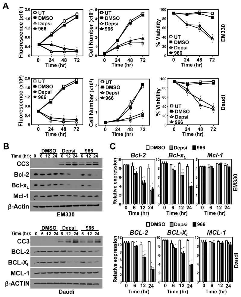
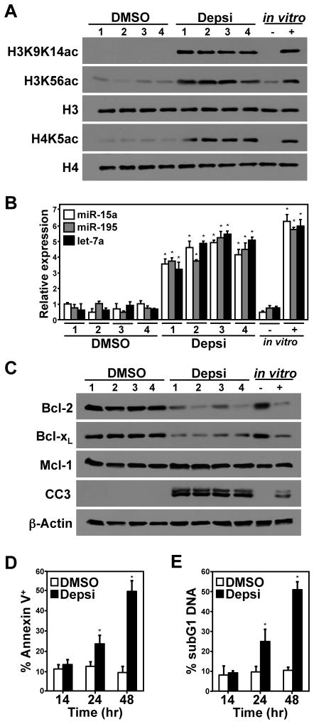
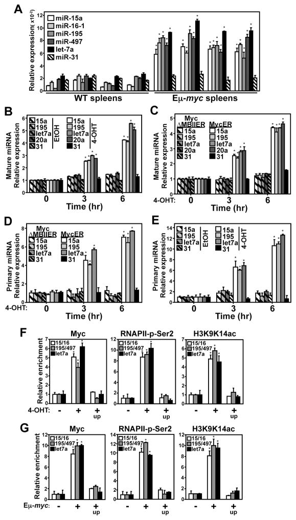
Alterations in the expression or function of histone deacetylases (HDAC) contribute to the development and progression of hematologic malignancies. Consequently, the development and implementation of HDAC inhibitors has proven to be therapeutically beneficial, particularly for hematologic malignancies. However, the molecular mechanisms by which HDAC inhibition (HDACi) induces tumor cell death remain unresolved. Here, we investigated the effects of HDACi in Myc-driven B-cell lymphoma and five other hematopoietic malignancies. We determined that Myc-mediated transcriptional repression of the miR-15 and let-7 families in malignant cells was relieved upon HDACi, and Myc was required for their upregulation. The miR-15 and let-7 families then targeted and downregulated the antiapoptotic genes Bcl-2 and Bcl-xL, respectively, to induce HDACi-mediated apoptosis. Notably, Myc also transcriptionally upregulated these miRNA in untransformed cells, indicating that this Myc-induced miRNA-mediated apoptotic pathway is suppressed in malignant cells, but becomes reactivated upon HDACi. Taken together, our results reveal a previously unknown mechanism by which Myc induces apoptosis independent of the p53 pathway and as a response to HDACi in malignant hematopoietic cells.


Conflict of interest statement

Conflict of Interest: The authors declare no conflict of interest.

Figures

Figure 1
Figure 1. HDACi reduces Bcl-2 and Bcl-xL expression and induces apoptosis
Cells remained untreated (UT) or received Depsipeptide (Depsi), RGFP966 (966), or vehicle (DMSO) control. (A) Following drug administration, proliferation (Alamar Blue; quadruplicate), cell number (triplicate), and viability (triplicate) were determined at the indicated intervals in murine (Eμ-myc, EM330) and human (Daudi) lymphoma cells. (B, C) Protein and mRNA levels were evaluated by Western blot (B) and qRT-PCR (triplicate) (C), respectively. Cleaved Caspase-3 (CC3). mRNA expression was normalized to β-Actin. Error bars are SD for A (*p<0.01) and SEM for C (*p<0.03); p-values determined by comparison to DMSO.
Figure 2
Figure 2. HDACi reverses Myc-mediated repression of the miR-15 family and let-7a
Lymphoma cells were treated with Depsipeptide (Depsi), RGFP966 (966), or vehicle (DMSO) control. (A) Western blots in murine (EM330) and human (Daudi) lymphoma cells were performed. (B, C) Mature miRNA levels were determined by qRT-PCR (triplicate) in (B) human diffuse large B-cell lymphoma (DLBCL, Burkitt’s lymphoma), two normal lymph nodes, and purified B-cells from peripheral blood (PB) and spleen (Sp), and (C) six murine Eμ-myc lymphomas, two pre-cancerous Eμ-myc spleens, and purified splenic B-cells. (D, E) Lymphoma cells were treated with Depsipeptide (Depsi), RGFP966 (966), or vehicle (DMSO) for the time indicated. Mature miRNA (D) or pri-miRNA (E) levels were determined by qRT-PCR (triplicate) in Eμ-myc (EM330) and Daudi lymphoma cells. Small RNA, RNU6b, was used for qRT-PCR normalization for B-E. (F) After treatment with Depsi or vehicle (DMSO) for 4hrs, ChIP with anti-RNAPII-phosphorylated-Serine2 (RNAPII-p-Ser2) or isotype control (IgG) was performed followed by qRT-PCR (triplicate) for the indicated promoters or upstream regions (up; negative controls) in murine (EM330) and human (Daudi) lymphoma cells. Values are relative to their respective IgG control and input DNA. (G) EM330 lymphoma cells were infected with either empty retrovirus (vector) or retrovirus encoding the miR-15a/16-1 or let-7a/7f miRNA clusters (14). Western blotting (left) was performed; U, uninfected; CC3, Cleaved Caspase-3. Viability (center; triplicate) and the percentage of cells with sub-G1 DNA content (right; triplicate) were determined at the indicated intervals. Error bars are SEM for B-F. B and C, *p<0.01, lymphoma versus mean of all normals; D-F, *p<0.001, Depsi or 966 versus DMSO. Error bars are SD for G, *p<0.03, compared to empty vector.
Figure 3
Figure 3. In vivo, HDACi increases miR-15 family and let-7a levels, inducing lymphoma cell death
C57Bl/6 mice with subcutaneous Eμ-myc lymphoma tumors (EM330) that reached 200mm3, were treated with Depsipeptide (Depsi) or vehicle (DMSO) control (n=4/group). Tumors were harvested 24hrs later and levels of the indicated proteins (A) and histone acetylation marks (C) were determined by Western blot. (B) miRNA levels were assessed by qRT-PCR (triplicate), and RNU6b was used for qRT-PCR normalization. As a positive control, cultured EM330 lymphoma cells (in vitro) were treated with vehicle (DMSO; -) or Depsi (+). Apoptosis was measured by cleaved Caspase-3 (CC3), AnnexinV-positivity (triplicate) (D), and propidium iodide staining of sub-G1 (apoptotic) DNA (triplicate) (E). Error bars are SEM for B (*p<0.001, Depsi versus mean of all DMSO controls) and SD for D and E (*p<0.03, Depsi versus DMSO).
Figure 4
Figure 4. Myc transcriptional activity is necessary to induce the miR-15 family and let-7a in non-transformed cells
Mature miRNA (A–C) and pri-miRNA (D, E) levels were determined by qRT-PCR (triplicate) and are normalized to RNU6b levels. (A) Pre-cancerous splenocytes from Eμ-myc mice and wild-type (WT) non-transgenic littermates were evaluated (n=4/group). (B–E) MycER or MycΔMBII-ER was activated with 4-OHT or vehicle (EtOH) control at the indicated intervals in primary pre-B-cells (B, E) and MEFs (C, D). (F, G) Following ChIP with anti-Myc, anti-RNAPII-phosphorylated-Serine2 (RNAPII-p-Ser2), anti-H3K9K14ac, or isotype controls (IgG), qRT-PCR for the indicated promoters or regions upstream (up) Myc does not bind (negative controls) was performed (triplicate). (F) MycER-expressing p53−/− MEFs received vehicle (EtOH; -) or 4-OHT (+) for 4hrs to induce MycER. (G) Splenocytes from wild-type (non-transgenic; Tg-) or pre-cancerous Eμ-myc transgenic (Tg+) littermate mice. Values for qChIP are relative to their respective IgG control and input DNA. Error bars are SEM; *p<0.001; (A) Eμ-myc versus mean of all WT spleens; (B, E, F) 4-OHT versus EtOH; (C, D) MycER versus MycΔMBII-ER, and (G) Eμ-myc (+) versus wild-type (−).
Figure 5
Figure 5. Myc is required for HDACi-induced transcriptional up-regulation of the miR-15 family and let-7a
(A, B, D, E) Following ChIP with anti-Myc, anti-RNAPII-phosphorylated-Serine2 (RNAPII-p-Ser2), anti-H3K9K14ac, or isotype controls (IgG), qRT-PCR for the indicated promoters or regions upstream (up) Myc does not bind (negative controls) was performed (triplicate). (A) Murine (EM330) and human (Daudi) lymphoma cells treated with Depsipeptide (Depsi) or vehicle (DMSO) for 4hrs. (B) p21 (Myc repression target) and CAD (Myc activation target) were controls (11). Values for qChIP are relative to their respective IgG control and input DNA. (C) Human P493-6 lymphoma cells containing tetracycline-regulatable MYC (P493-6) exposed to tetracycline (+; MYC-OFF) for 24hrs or not (MYC-ON) were Western blotted. These cells were also treated for 12hrs with Depsi or vehicle (DMSO) control. miRNA were measured by qRT-PCR (triplicate) and normalized to RNU6b levels. (D, E) Before qChIP analyses, P493-6 cells received tetracycline for 24hrs (+; MYC-OFF) or not (−; MYC-ON) prior to treatment with Depsipeptide (Depsi) or vehicle (DMSO) for 4hrs. Values for qChIP are relative to their respective IgG control and input DNA. Error bars are SEM; *p<0.0002 (B), *p<0.01 (E), both RNAPII-p-Ser2 and H3K9K14ac versus IgG (B), Depsi versus DMSO (C).
Figure 6
Figure 6. Myc transcriptional activity regulates Bcl-2 and Bcl-xL expression independent of Miz-1 and p53
(A) Eμ-myc lymphomas (n=11) and pre-cancerous Eμ-myc purified B-cells (n=2) and spleens (n=3) were Western blotted. (B) Pre-cancerous splenocytes from Eμ-myc mice (n=5) and wild-type (WT) non-transgenic littermates (n=5) were Western blotted. (C, D, F) At intervals following addition of 4-OHT, wild-type (WT) MycER-expressing murine embryonic fibroblasts (MEFs) and primary pre-B-cells (C) and MycV394D-ER- (D) and MycΔMBII-ER-expressing (F) WT MEFs were harvested and Western blotted. (E) miRNA levels were determined by qRT-PCR (triplicate; normalized to RNU6b levels) following MycV394D-ER activation with 4-OHT or vehicle (EtOH). Error bars are SEM; *p<0.001, 4-OHT versus EtOH.
Figure 7
Figure 7. Myc induces the miR-15 family and let-7a that then target Bcl-2 and Bcl-xL
(A, C) Luciferase expression vectors containing the 3′-untranslated region (3′-UTR) of Bcl-2 and Bcl-xL with the wild-type (WT) or a mutated (Mut) miRNA binding site were transfected into fibroblasts expressing the 4-OHT-inducible MycER or MycΔMBII-ER. An expression vector containing the miR-17 family binding site of the wild-type (WT) p21 3′-UTR was a positive control (13). Luciferase activity was measured (triplicate) 48hrs following vehicle (EtOH) control or 4-OHT addition to activate MycER. A β-galactosidase reporter plasmid was co-transfected for normalization. (B–F) miR-15 family and let-7a miRNA binding sites in the Bcl-2 and Bcl-xL 3′-UTR were blocked with site-specific small molecules (TP; Target Protectors). (B) Wild-type MEFs transfected with either Bcl-2 or Bcl-xL Target Protectors (TP) and/or miR-15a mimic were Western blotted. UT, untransfected cells. (D–F) p53−/− MEFs, with or without Bcl-2 and/or Bcl-xL Target Protectors (TP), expressing the 4-OHT-inducible MycV394D-ER were Western blotted (D), subjected to MTT assay (E; quadruplicate), or analyzed for AnnexinV-positivity (triplicate) by flow cytometry at intervals following addition of 4-OHT. Cleaved Caspase-3 (CC3). Error bars are SEM (A and C, *p<0.009, 4-OHT versus EtOH) and SD (E, *p<0.02; F, *p<0.0001; both TP versus control).

References

    1. West AC, Johnstone RW. New and emerging HDAC inhibitors for cancer treatment. J Clin Invest. 2014;124(1):30–9. - PMC - PubMed
    1. Conti C, Leo E, Eichler GS, Sordet O, Martin MM, Fan A, et al. Inhibition of histone deacetylase in cancer cells slows down replication forks, activates dormant origins, and induces DNA damage. Cancer research. 2010;70(11):4470–80. - PMC - PubMed
    1. Stengel KR, Hiebert SW. Antioxidants & redox signaling. 2014. Class I HDACs Affect DNA Replication, Repair, and Chromatin Structure: Implications for Cancer Therapy. - PMC - PubMed
    1. Dang CV. MYC on the path to cancer. Cell. 2012;149(1):22–35. - PMC - PubMed
    1. Eischen CM, Weber JD, Roussel MF, Sherr CJ, Cleveland JL. Disruption of the ARF-Mdm2-p53 tumor suppressor pathway in Myc-induced lymphomagenesis. Genes & development. 1999;13(20):2658–69. - PMC - PubMed

Publication types

Substances