. 2015 Dec 16;90(5):2455-72.
doi: 10.1128/JVI.02942-15.

Deletion of Murid Herpesvirus 4 ORF63 Affects the Trafficking of Incoming Capsids toward the Nucleus

Affiliations

Deletion of Murid Herpesvirus 4 ORF63 Affects the Trafficking of Incoming Capsids toward the Nucleus

Muhammad Bilal Latif et al. J Virol. .

Abstract

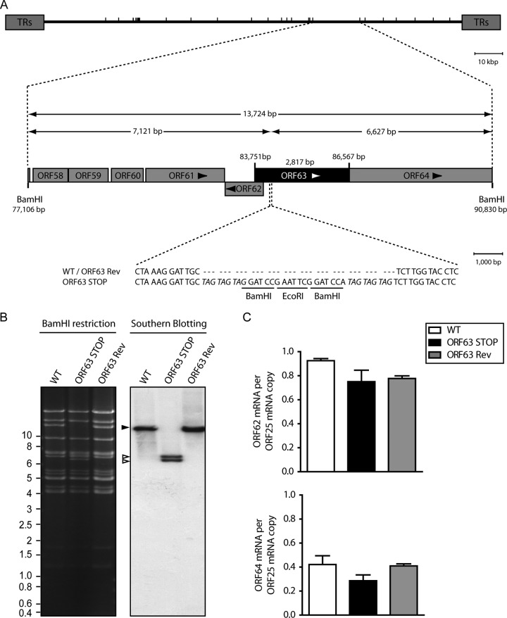
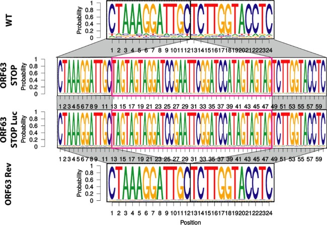
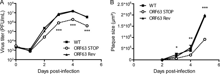
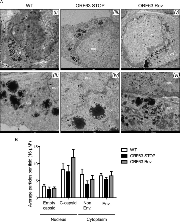
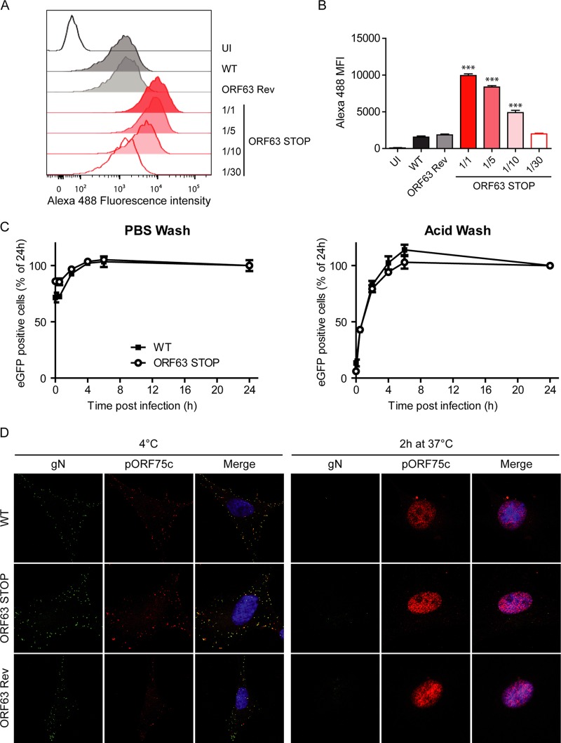
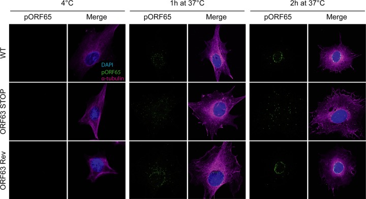
Gammaherpesviruses are important human and animal pathogens. Despite the fact that they display the classical architecture of herpesviruses, the function of most of their structural proteins is still poorly defined. This is especially true for tegument proteins. Interestingly, a potential role in immune evasion has recently been proposed for the tegument protein encoded by Kaposi's sarcoma-associated herpesvirus open reading frame 63 (ORF63). To gain insight about the roles of ORF63 in the life cycle of a gammaherpesvirus, we generated null mutations in the ORF63 gene of murid herpesvirus 4 (MuHV-4). We showed that disruption of ORF63 was associated with a severe MuHV-4 growth deficit both in vitro and in vivo. The latter deficit was mainly associated with a defect of replication in the lung but did not affect the establishment of latency in the spleen. From a functional point of view, inhibition of caspase-1 or the inflammasome did not restore the growth of the ORF63-deficient mutant, suggesting that the observed deficit was not associated with the immune evasion mechanism identified previously. Moreover, this growth deficit was also not associated with a defect in virion egress from the infected cells. In contrast, it appeared that MuHV-4 ORF63-deficient mutants failed to address most of their capsids to the nucleus during entry into the host cell, suggesting that ORF63 plays a role in capsid movement. In the future, ORF63 could therefore be considered a target to block gammaherpesvirus infection at a very early stage of the infection.

Importance: The important diseases caused by gammaherpesviruses in human and animal populations justify a better understanding of their life cycle. In particular, the role of most of their tegument proteins is still largely unknown. In this study, we used murid herpesvirus 4, a gammaherpesvirus infecting mice, to decipher the role of the protein encoded by the viral ORF63 gene. We showed that the absence of this protein is associated with a severe growth deficit both in vitro and in vivo that was mainly due to impaired migration of viral capsids toward the nucleus during entry. Together, our results provide new insights about the life cycle of gammaherpesviruses and could allow the development of new antiviral strategies aimed at blocking gammaherpesvirus infection at the very early stages.


Figures

FIG 1
FIG 1
Comparison of MuHV-4 ORF63 with its homologs in other herpesviruses. Shown is a multiple-sequence alignment of the protein encoded by MuHV-4 ORF63 with its homologs in different herpesviruses: HSV-1 UL37 (GI 692148201), HCMV UL47 (GI 44903273), EBV BOLF1 (GI 764007616), and KSHV ORF63 (GI 139472855). Αlpha-helices are highlighted in red boxes, while beta-sheets are highlighted in blue. The conservation at individual amino acids positions is shown below the sequences. Conservation in all the strains is highlighted in red.
FIG 2
FIG 2
Generation of a ORF63-deficient MuHV-4 mutant. (A) Schematic representation of the strategy followed to produce the recombinant MuHV-4 strains. The ORF63-deficient MuHV-4 mutant was derived from a cloned MuHV-4 BAC by a galK counterselection method. The ORF63 coding sequence was disrupted by inserting stop codons (ORF63 STOP). The mutation incorporated new BamHI restriction sites. This virus was reverted by homologous recombination with a WT genomic segment (ORF63 Rev). TRs, terminal repeats. (B) Verification of the molecular structure. BAC DNA was digested with BamHI, resolved by agarose gel electrophoresis, and hybridized with a 32P-labeled probe, corresponding to nucleotides 83819 to 84693 of the MuHV-4 WUMS strain genome. The black arrowhead shows the WT ORF63 fragment (13,724 bp). Open arrowheads show the restriction fragments that contain ORF63 STOP (7,121 bp and 6,627 bp, respectively, for the left and the right fragments). Sizes in kilobase pairs are indicated on the left. (C) Viral transcription in ORF63+ and ORF63 viruses. BHK-21 cells were infected with WT, ORF63 STOP, and ORF63 Rev MuHV-4 strains (0.5 PFU/cell), and 24 h later, RNA was extracted, reverse transcribed, and assayed for viral transcripts by qRT-PCR amplification of part of each gene. ORF62 and ORF64 flank the ORF63 gene of MuHV-4. ORF25 is a control viral gene. The data are averages from triplicate measurements ± SEMs and were analyzed by 1-way analysis of variance (ANOVA) and Bonferroni posttests.
FIG 3
FIG 3
Verification of the molecular structures of the viral genomes after in vitro growth. Shown is a schematic representation as a sequence logo of the aligned sequences of the region encompassing the insertion point of the stop codons for the WT, ORF63 STOP, ORF63 STOP Luc, and ORF63 Rev strains. These sequencing reactions have been performed on viral DNA from purified virions. All the genomes displayed the expected molecular structures.
FIG 4
FIG 4
In vitro effect of the ORF63 deficiency on growth of MuHV-4. (A) Effect of ORF63 deficiency on MuHV-4 growth in vitro. BHK-21 cells were infected with WT, ORF63 STOP, and ORF63 Rev MuHV-4 strains in 6-well cluster dishes at an MOI of 0.01 PFU per cell. Supernatant and infected cells were harvested at different times after infection, and the amount of infectious virus was determined by plaque assay on BHK-21 cells. The data are averages from triplicate measurements ± SEMs and were analyzed by 2-way ANOVA and Bonferroni posttests. ***, P < 0.001. At time zero p.i., the inocula were retitrated to ensure that similar amounts of virus were put on the cells. (B) Effect of the absence of pORF63 on MuHV-4 plaque size. BHK-21 cells grown on coverslips were infected with MuHV-4 WT, ORF63 STOP, and ORF63 Rev strains and then overlaid with DMEM containing CMC as described in Materials and Methods. At successive intervals after infection, plaques were fixed and measured. Each datum point is the average ± SEM for the measurement of 20 plaques per time point. The data were analyzed by 2-way ANOVA and Bonferroni posttests. *, P < 0.05; **, P < 0.01; ***, P < 0.001.
FIG 5
FIG 5
Generation of an ORF63-deficient MuHV-4 mutant expressing luciferase. (A) Schematic representation of the strategy followed to produce the recombinant MuHV-4 Luc strains. The ORF63 STOP Luc strain was derived from a cloned MuHV-4 BAC-Luc strain by a galK counterselection method. The ORF63 coding sequence was disrupted by inserting stop codons (ORF63 STOP). The mutation incorporated new BamHI restriction sites. This virus was reverted by homologous recombination with an unmutated genomic segment (ORF63 Rev). (B) Verification of the molecular structure. BAC DNA was digested with BamHI, resolved by agarose gel electrophoresis, and hybridized with a 32P-labeled probe corresponding to nucleotides 83819 to 84693 of the MuHV-4 WUMS strain genome. The black arrowhead shows WT ORF63 fragment (13,649 bp). Open arrowheads show the restriction fragments that contain ORF63 STOP (7,046 bp and 6,627 bp, respectively, for the left and the right fragments). Sizes in kilobase pairs are indicated on the left. (C) Viral transcription in ORF63+ and ORF63 Luc viruses. BHK-21 cells were infected with WT Luc, ORF63 STOP Luc, and ORF63 Rev Luc MuHV-4 strains (0.5 PFU/cell), and 24 h later RNA was extracted, reverse transcribed, and assayed for viral transcripts by qRT-PCR amplification of part of each gene. ORF62 and ORF64 flank the ORF63 gene of MuHV-4. ORF25 is a control viral gene. The data are averages from triplicate measurements ± SEMs and were analyzed by 1-way ANOVA and Bonferroni posttests. (D) Effect of ORF63 deficiency on in vitro growth of MuHV-4 strains expressing Luc. BHK-21 cells were infected with WT Luc, ORF63 STOP Luc, and ORF63 Rev Luc MuHV-4 strains in 6-well cluster dishes at an MOI of 0.01 PFU per cell. Supernatant and infected cells were harvested at different times after infection, and the amount of infectious virus was determined by plaque assay on BHK-21 cells. The data are averages from triplicate measurements ± SEMs and were analyzed by 2-way ANOVA and Bonferroni posttests; ***, P < 0.001. At time zero p.i., the inocula were retitrated to ensure that similar amounts of virus were put on the cells.
FIG 6
FIG 6
In vivo infection by luciferase-expressing MuHV-4 strains. Female mice were infected intranasally (1 × 10⁴ PFU) with MuHV-4 WT Luc (top row) or ORF63 STOP Luc (bottom rpw) strains under general anesthesia. The mice were then injected with luciferin and imaged at the indicated time points. Images show a representative mouse (from a group of 5 mice) at days 3, 5, 7, 10, 12, 14, 17, and 19 p.i. The scale bar shows the color scheme for signal intensity.
FIG 7
FIG 7
Effect of ORF63 deficiency on replication of MuHV-4 in vivo. (A) BALB/c mice were infected intranasally with WT, ORF63 STOP, and ORF63 Rev MuHV-4 strains (1 × 10⁴ PFU). At the indicated times p.i., the infectious virus titers in lungs were determined by plaque assay. (B) Individual sera collected at the different time points were analyzed for MuHV-4-specific IgG by ELISA. Pooled naive sera provided the negative control. The data are averages from 5 mice ± SEMs and were analyzed by 2-way ANOVA and Bonferroni posttests. ***, P < 0.001; **, P < 0.01. (C) Lung histology. Seven days after infection with the different strains of MuHV-4, lungs were removed and fixed in formaldehyde before hematoxylin-eosin staining. Rectangles identify regions that are highlighted in higher-magnification pictures. Arrows indicate perivascular and peribronchial lymphocyte accumulation. The images are representative of data from at least 5 animals. (D) Spleens from the same mice were analyzed individually by infectious-center assay. (E) DNA was extracted from individual spleens. The viral genome copy number was then determined by real-time PCR.
FIG 8
FIG 8
The growth deficit of the MuHV-4 ORF63 STOP mutant strain is not associated with an increased cell death or with the activation of the inflammasome. (A) Kinetic of ORF63 expression. BHK-21 cells were infected with MuHV-4 (MOI of 0.5 PFU/cell). At the indicated time postinfection, expression of ORF63 was studied by a Sybr green qRT-PCR approach as described in Materials and Methods. Time zero represents uninfected cells. The data are averages from triplicate measurements ± SEMs and were analyzed by 1-way ANOVA and Bonferroni posttests. ***, P < 0.001. (B) BHK-21 cells were mock infected or infected with WT BAC+, ORF63 STOP BAC+, and ORF63 Rev BAC+ MuHV-4 strains at an MOI of 0.5 PFU/cell. Twenty-four hours after infection, cell viability was assessed by annexin V-APC and propidium iodide labeling and flow cytometry analysis. Percentages of doubly positive cells were measured in eGFP and eGFP+ populations. The data are averages from triplicates ± SEMs and were analyzed by 2-way ANOVA and Bonferroni posttests. (C and D) Effect of Z-VAD (C) or glyburide (D) on growth of MuHV-4 in vitro. BHK-21 cells were infected with WT, ORF63 STOP, and ORF63 Rev MuHV-4 strains in 6-well cluster dishes at an MOI of 0.01 PFU per cell in the presence of the pan-caspase inhibitor Z-VAD (20 μM) or the NLRP3 inflammasome inhibitor glyburide (25 μg/ml). Supernatant and infected cells were harvested at different times after infection, and the amounts of infectious virus were determined by plaque assay on BHK-21 cells. The data are averages from triplicate measurements ± SEMs and were analyzed by 2-way ANOVA and Bonferroni posttests. **, P < 0.01; ***, P < 0.001. At time zero p.i., the inocula were retitrated to ensure that similar amounts of virus were put on the cells. (E) Effect of infection supernatant on the growth of MuHV-4 in vitro. BHK-21 cells were infected with WT, ORF63 STOP, and ORF63 Rev MuHV-4 strains in 24-well cluster dishes at an MOI of 0.01 PFU per cell in the presence of supernatant of BHK-21 cells previously infected with WT, ORF63 STOP, and ORF63 Rev MuHV-4 strains (500 μl/well; 50% final concentration). Supernatant and infected cells were harvested at different times after infection and the amounts of infectious virus were determined by plaque assay on BHK-21 cells. The data are averages from triplicate measurements ± SEMs and were analyzed by 2-way ANOVA and Bonferroni posttests. **, P < 0.01; ***, P < 0.001. At time zero p.i., the inocula were retitrated to ensure that similar amounts of virus were put on the cells. To allow comparisons between graphs, a dashed line has been added across the graphs at the mean maximal value measured for WT and ORF63 Rev strains.
FIG 9
FIG 9
ORF63 deficiency is not associated with a morphogenesis defect or an egress deficit but is associated with an increased particle/PFU ratio. (A) Transmission electron microscopic (TEM) analysis for morphogenesis of the virions. BHK-21 cells were infected with WT (i and ii), ORF63 STOP (iii and iv), and ORF63 Rev (v and vi) MuHV-4 strains (1 PFU/cell, 48 h), washed with PBS, and fixed in TEM fixation buffer for TEM. The scale bars are shown below the images. nuc, nucleus; cyt, cytoplasm. No difference was observed in the assembly of nucleocapsids and their transport from the nucleus (black arrows; approximate diameter, 100 nm) among the infected cells by different viral strains. The enveloped viruses are shown with the white arrows in the cytoplasms of the infected cells. No difference in virus egress was found among the samples. (B) Distribution of different virus capsids and particle types in TEM micrographs of BHK-21 cells that were infected with the WT, ORF63 STOP, or ORF63 Rev strains for 48 h at an MOI of 0.5 PFU/cell. Average numbers of particles from 6 to 9 different micrographs spanning at least 3 different infected cells for each sample were identified based on their established characteristics (61) and enumerated before statistical analysis was performed (GraphPad software).
FIG 10
FIG 10
ORF63 deficiency is associated with an increased particle/PFU ratio and a deficit in entry. (A) High-MOI growth curve. BHK-21 cells were infected with WT, ORF63 STOP, and ORF63 Rev MuHV-4 strains in 6-well cluster dishes at an MOI of 1 PFU per cell. Supernatant and infected cells were harvested independently at different times after infection, and the amounts of infectious virus were determined for both kinds of samples by plaque assay on BHK-21 cells. The data are the averages from triplicate measurements ± SEMs and were analyzed by 2-way ANOVA and Bonferroni posttests. ***, P < 0.001. At time zero p.i., the inocula were retitrated to ensure that similar amounts of virus were put on the cells. (B) Comparison of the structural proteins content for different amounts of PFU (3 × 10⁴, 1 × 10⁴, and 3 × 10³) between the different strains. MuHV-4 WT, ORF63 STOP, and ORF63 Rev stocks were compared for viral protein content by immunoblotting with anti-MuHV-4 rabbit polyserum. (C) A total of 106 BHK-21 cells were infected with WT, ORF63 STOP, and ORF63 Rev MuHV-4 strains at an MOI of 0.5 PFU/cell. For the ORF63 STOP strain, an additional sample of cells infected by a number of particles equivalent to the WT and ORF63 Rev strains was added. Six hours later, RNA was extracted, reverse transcribed, and assayed for ORF50 expression by qPCR amplification. The data are averages from triplicate measurements ± SEMs and were analyzed by 1-way ANOVA and Bonferroni posttests. (D) BHK-21 cells were exposed to eGFP expressing (BAC+) WT, ORF63 STOP, and ORF63 Rev strains (0.5 PFU/cell). For the ORF63 STOP strain, an additional sample of cells infected by a number of particles equivalent to those of the WT and ORF63 Rev strains (determined by Western blotting) was added. After binding for the times indicated, the cells were washed with PBS and assayed by flow cytometry for eGFP expression. The data are the averages ± SEMs from triplicate measurements. The data were analyzed by 2-way ANOVA and Bonferroni posttests. ***, P < 0.001.
FIG 11
FIG 11
Binding, endocytosis, and fusion of ORF63 STOP virions. (A and B) WT, ORF63 STOP, and ORF63 Rev virions were bound to BHK-21 cells (3 h, 4°C) with either the same numbers of PFU for the three strains or dilutions of this amount (1/5, 1/10, 1/30) for the ORF63 STOP strain. Cell surface-bound virions were detected by washing, fixing, and staining them for gN with MAb 3F7 (38). Secondary detection was with Alexa 488-conjugated goat anti-mouse IgG pAb. The cells were then analyzed by flow cytometry (A). This experiment was performed in triplicates (B), and the differences in gN detection (mean fluorescence intensity [MFI]) between samples were analyzed by 1-way ANOVA and Bonferroni posttests. ***, P < 0.001 compared to the value for the WT sample. (C) BHK-21 cells were exposed to WT, ORF63 STOP, and ORF63 Rev BAC+ (expressing eGFP) strains (0.5 PFU/cell) for the times indicated and then washed either with PBS (pH 7.4) or with isotonic buffer (pH 3; acid wash). Viral infection was assayed by measuring eGFP expression 24 h p.i. by flow cytometry. The data are average ± SEMs for triplicate measurements. The data were analyzed by 2-way ANOVA and Bonferroni posttests. (D) MuHV-4 WT, ORF63 STOP, and ORF63 Rev virions were bound to BHK-21 cells (particles equivalent to 30 WT PFU/cell, 3 h, 4°C). The cells were then washed with PBS and either fixed immediately or first further incubated (2 h, 37°C) to allow virion endocytosis and membrane fusion. The cells were then stained for the gN envelope glycoprotein (IgG2a [green]) and for the ORF75c virion tegument protein with MAb BN-8C3 (IgG1 [red]), and with DAPI (blue). Red and green colocalization appears as yellow. Equivalent data were obtained in a repeat experiment. The data are fully representative of at least 100 cells examined. The confocal settings were the same for the corresponding images at 4°C and after 2 h at 37°C.
FIG 12
FIG 12
Effect of ORF63 deficiency on capsid localization during entry. MuHV-4 WT, ORF63 STOP, and ORF63 Rev virions were bound to BHK-21 cells (particles equivalent to 30 WT PFU/cell, 3 h, 4°C). The cells were then washed with PBS and either fixed immediately or first further incubated (1 or 2 h, 37°C) to allow virion endocytosis, membrane fusion, and capsid release into the cytoplasm. The cells were then stained for the ORF65 capsid protein with MAb 12B8 (IgG2a [green]; epitope only revealed after fusion) and for the alpha-tubulin (rat IgG [magenta]), and with DAPI (blue). Equivalent data were obtained in a repeat experiment. The data shown are fully representative of those obtained for at least 100 cells examined. The confocal settings were the same for the corresponding images at 4°C and after 1 or 2 h at 37°C.

References

    1. Ackermann M. 2006. Pathogenesis of gammaherpesvirus infections. Vet Microbiol 113:211–222. doi: 10.1016/j.vetmic.2005.11.008. - DOI - PubMed
    1. Thorley-Lawson DA, Gross A. 2004. Persistence of the Epstein-Barr virus and the origins of associated lymphomas. N Engl J Med 350:1328–1337. doi: 10.1056/NEJMra032015. - DOI - PubMed
    1. Verma SC, Robertson ES. 2003. Molecular biology and pathogenesis of Kaposi sarcoma-associated herpesvirus. FEMS Microbiol Lett 222:155–163. doi: 10.1016/S0378-1097(03)00261-1. - DOI - PubMed
    1. Henle G, Henle W, Clifford P, Diehl V, Kafuko GW, Kirya BG, Klein G, Morrow RH, Munube GM, Pike P, Tukei PM, Ziegler JL. 1969. Antibodies to Epstein-Barr virus in Burkitt's lymphoma and control groups. J Natl Cancer Inst 43:1147–1157. - PubMed
    1. Damania B. 2004. Oncogenic gamma-herpesviruses: comparison of viral proteins involved in tumorigenesis. Nat Rev Microbiol 2:656–668. doi: 10.1038/nrmicro958. - DOI - PubMed

Publication types

LinkOut - more resources