. 2015 Dec 16;90(5):2586-99.
doi: 10.1128/JVI.02420-15.

Mouse Hepatitis Virus Infection Remodels Connexin43-Mediated Gap Junction Intercellular Communication In Vitro and In Vivo

Affiliations

Mouse Hepatitis Virus Infection Remodels Connexin43-Mediated Gap Junction Intercellular Communication In Vitro and In Vivo

Rahul Basu et al. J Virol. .

Abstract

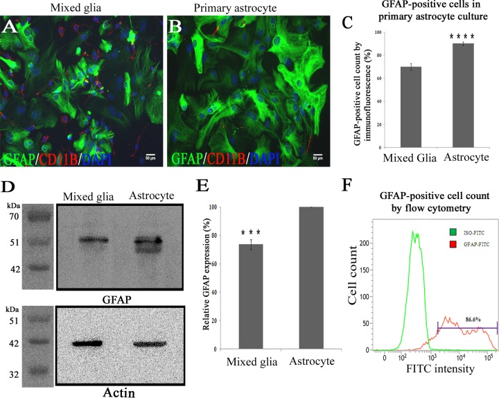
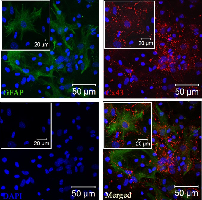
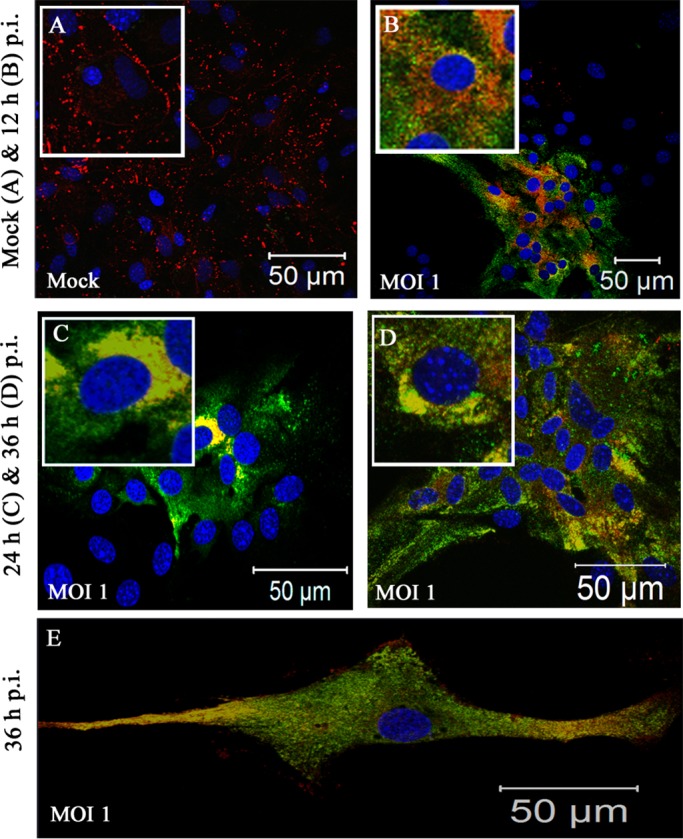
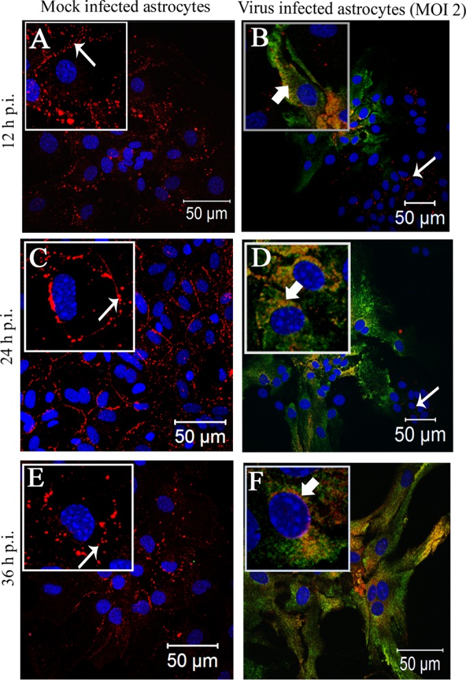
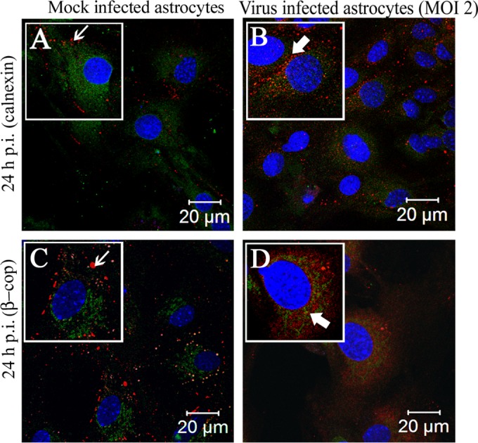
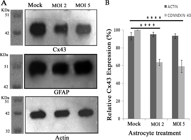
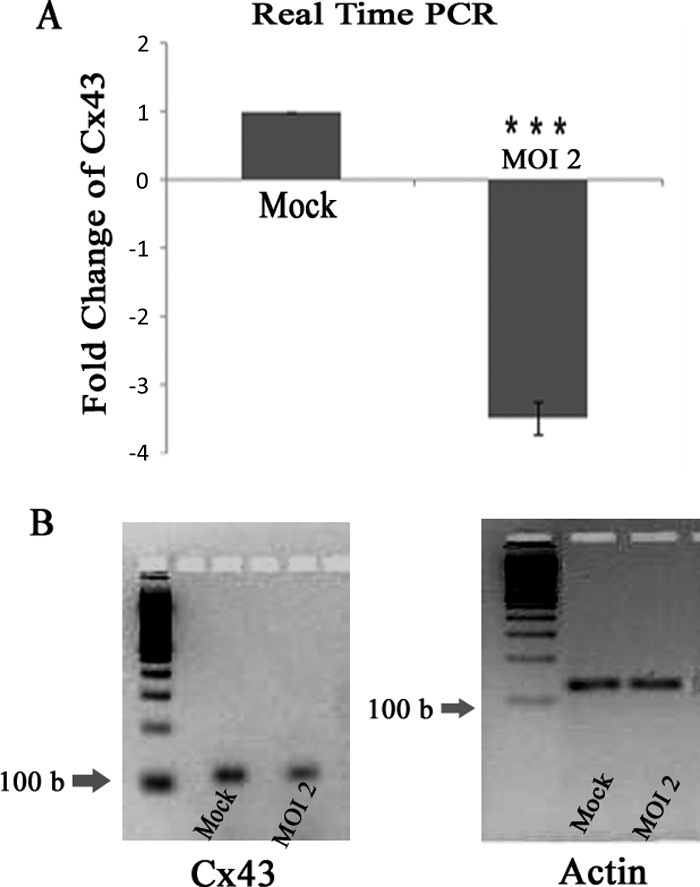
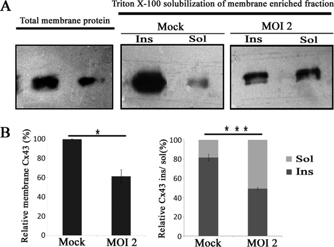
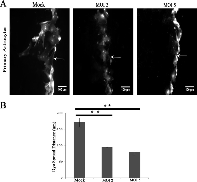
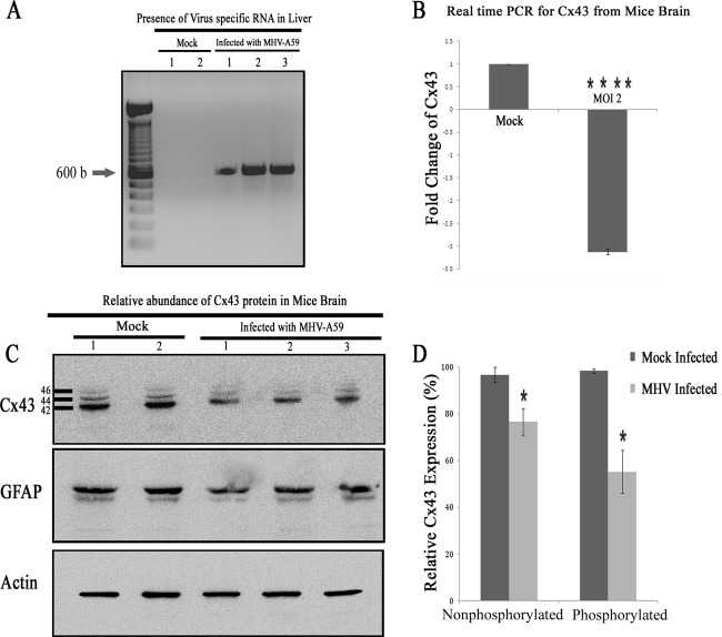
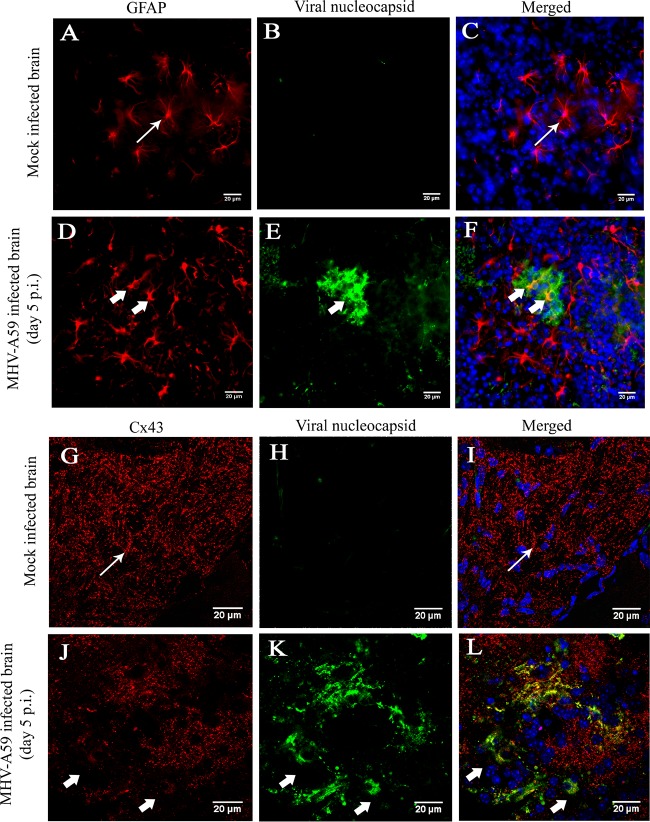
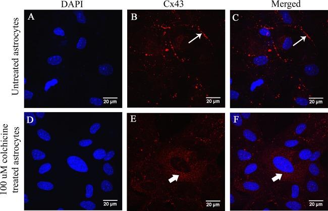
Gap junctions (GJs) form intercellular channels which directly connect the cytoplasm between neighboring cells to facilitate the transfer of ions and small molecules. GJs play a major role in the pathogenesis of infection-associated inflammation. Mutations of gap junction proteins, connexins (Cxs), cause dysmyelination and leukoencephalopathy. In multiple sclerosis (MS) patients and its animal model experimental autoimmune encephalitis (EAE), Cx43 was shown to be modulated in the central nervous system (CNS). The mechanism behind Cx43 alteration and its role in MS remains unexplored. Mouse hepatitis virus (MHV) infection-induced demyelination is one of the best-studied experimental animal models for MS. Our studies demonstrated that MHV infection downregulated Cx43 expression at protein and mRNA levels in vitro in primary astrocytes obtained from neonatal mouse brains. After infection, a significant amount of Cx43 was retained in endoplasmic reticulum/endoplasmic reticulum Golgi intermediate complex (ER/ERGIC) and GJ plaque formation was impaired at the cell surface, as evidenced by a reduction of the Triton X-100 insoluble fraction of Cx43. Altered trafficking and impairment of GJ plaque formation may cause the loss of functional channel formation in MHV-infected primary astrocytes, as demonstrated by a reduced number of dye-coupled cells after a scrape-loading Lucifer yellow dye transfer assay. Upon MHV infection, a significant downregulation of Cx43 was observed in the virus-infected mouse brain. This study demonstrates that astrocytic Cx43 expression and function can be modulated due to virus stress and can be an appropriate model to understand the basis of cellular mechanisms involved in the alteration of gap junction intercellular communication (GJIC) in CNS neuroinflammation.

Importance: We found that MHV infection leads to the downregulation of Cx43 in vivo in the CNS. In addition, results show that MHV infection impairs Cx43 expression in addition to gap junction communication in primary astrocytes. After infection, Cx43 did not traffic normally to the membrane to form gap junction plaques, and that could be the basis of reduced functional gap junction coupling between astrocytes. This is an important first step toward understanding how viruses affect Cx43 expression and trafficking at the cellular level. This may provide a basis for understanding how structural alterations of astrocytic gap junctions can disrupt gap junction communication between other CNS cells in altered CNS environments due to infection and inflammation. More specifically, alteration of Cx43 may be the basis of the destabilization of Cx47 in oligodendrocytes seen in and around inflammatory demyelinating plaques in MS patients.


Figures

FIG 1
FIG 1
Enrichment and characterization of astrocytes enriched from mixed glial culture. One confluent monolayer of mixed glial cells (A) and an astrocyte-enriched primary culture (B) isolated from neonatal mouse brain were labeled with anti-GFAP antibody (astrocytic marker; green) and CD11b (microglia marker; red). Cells were counterstained with DAPI (nuclear stain; blue). (C) Visual counting of immunostained cells demonstrated that mixed glial cultures consist of GFAP+ astrocytes (70% ± 3.0%) and CD11b+ microglia (9% ± 2%), whereas astrocyte-enriched cultures demonstrated a significantly higher percentage (90.3% ± 1.5%) of cells that were positive for GFAP (data were mean values ± SEM from three experimental sets having 10 replicates each; ****, P < 0.0001). (D) Five micrograms of total protein from mixed glial cultures as well as astrocyte-enriched cultures was immunoblotted for GFAP and an internal control, γ-actin. (E) Astrocyte-enriched cultures demonstrated 26.3% ± 3.4% higher GFAP expression (normalized to internal control actin) than mixed glial cultures (data were mean values ± SEM from three experiment sets; ***, P < 0.001). (F) For quantification, primary astrocytes were immunostained with FITC-labeled anti-GFAP antibody and subjected to flow cytometric analysis. A total of 86.6% of cells were positive for GFAP (one representative plot of three experiments is shown).
FIG 2
FIG 2
Expression of Cx43 at the cell surface of GFAP+ primary astrocytes. Primary astrocytes were double immune labeled with mouse anti-GFAP (astrocyte marker) and rabbit anti-Cx43 antisera. Cells subsequently were labeled with FITC goat anti-mouse IgG and Texas Red goat anti-rabbit IgG, respectively. Immunostained cells were counterstained with DAPI (blue). Merged images show that GFAP-positive astrocytes express high levels of punctate Cx43 staining at the cell surface. Insets show magnifications of the expression of Cx43 at the cell surface of GFAP+ astrocytes.
FIG 3
FIG 3
Intracellular localization of Cx43 in MHV-A59-infected primary astrocytes at an MOI of 1. Primary astrocytes were mock infected (A) or infected with MHV-A59 at an MOI of 1 (B to E) and subjected to double-label immunofluorescence with anti-Cx43 antisera (red) and antiviral nucleocapsid (N) antisera (green). Astrocytes demonstrated heterogeneous morphology. At this low viral dose, syncytia were not found to be present, even at 36 h p.i. Infected cells showed characteristic intracellular retention of Cx43 upon viral infection. Astrocytes which were part of a confluent monolayer (B to D), as well as astrocytes which grew as isolated large single cells (E), showed similar retention of Cx43 in an intracellular compartment after infection.
FIG 4
FIG 4
Intracellular localization of Cx43 in MHV-A59-infected primary astrocytes at an MOI of 2. Primary astrocytes were mock infected (A, C, and E) or infected with MHV-A59 at an MOI of 2 (B, D, and F). Cells were subjected to double-label immunofluorescence at 12 h (A and B), 24 h (C and D), and 36 h (E and F) p.i. with anti-Cx43 antisera (red) and anti-N antisera (green). Cells were visualized at 40× on a laser-scanning microscope. (A, C, and E) For the mock-infected cells, Cx43 was localized at the cell surface (thin arrow) with very minimal distribution in the intracellular compartment. In contrast, in MHV-A59-infected cells Cx43 was localized primarily in the intracellular compartment with very minimal distribution at the cell surface and was mostly colocalized with anti-N antisera (B, D, and E, thick arrow). Interestingly, intracellular retention of Cx43 was restricted to infected cells only. Cells negative for viral-N (B and D, thin arrow) did not show retention of Cx43 in an intracellular compartment. At each time point p.i., mock-infected cells (A, C, and E) expressed Cx43 at the cell surface (thin arrow), whereas Cx43 was localized mainly in the perinuclear compartment (B, D, and F, thick arrow) and partially colocalized with viral N protein in infected cells. To better illustrate these observed localization patterns, a single cell from the confluent astrocyte monolayer is shown at higher magnification in an inset.
FIG 5
FIG 5
Localization of Cx43 predominantly in the ER/ERGIC of virus-infected cells. Primary astrocytes were mock infected or infected with MHV-A59 at an MOI of 2 and were subjected to double-label immunofluorescence at 24 h p.i. with anti-Cx43 antisera (red) and anti-calnexin (green) or anti-β-cop antisera(green). The images show prominent punctate staining of Cx43 at the cell surface (A and C, thin arrow), forming gap junction plaques, in mock-infected cells. Cx43 in the virus-infected cells, which was retained in the intracellular compartments, mostly colocalized with the ER marker calnexin and/or ERGIC marker β-cop (B and D, thick arrow).
FIG 6
FIG 6
Reduction of Cx43 protein expression due to MHV-A59 infection. Primary astrocytes were either mock infected or infected with MHV-A59, and total protein was extracted at 24 h p.i. Protein (5 μg) was resolved in SDS-PAGE, transferred to a PVDF membrane, and immunoprobed. (A) Upon infection with MHV-A59, whole-cell Cx43 protein levels were decreased compared to those of mock-infected cells. The internal loading control γ-actin showed similar signal intensity for all experiments. (B) Densitometric analysis showed that there was a 36.3% ± 3.3% (at an MOI of 2) and 40.8% ± 6.8% (at an MOI of 5) reduction in total Cx43 for MHV-infected cells compared to the level for mock-infected control cells. The total proteins isolated from primary astrocytes also were probed for GFAP, but expression was found to be similar in all experimental groups. The mean ± SEM incidences of six experimental replicates from three different experiments are shown (****, P < 0.0001).
FIG 7
FIG 7
Reduction of Cx43 mRNA abundance in viral infection. Primary astrocytes were infected with MHV-A59 at an MOI of 2, and mock-infected cells were maintained in parallel. RNA was extracted at 24 h p.i., and subsequently cDNA was synthesized. Equal amounts of cDNA template were used for qPCR analysis. The relative expression of Cx43 mRNA was obtained using the ΔΔCT method. (A) Compared to that of mock-infected cells, Cx43 expression was 3.49- ± 0.25-fold downregulated after MHV-A59 infection. The end products of the qPCR of Cx43 and an internal control, β-actin, were resolved in a 4% agarose gel and are shown in panel B. Mean ± SEM incidences from three different experiments are shown (***, P < 0.001).
FIG 8
FIG 8
Confirmation of reduced gap junction plaque formation due to infection using Triton X-100 solubilization. Primary astrocytes were infected with MHV-A59 at an MOI of 2, and mock-infected cells were maintained in parallel. A membrane-enriched fraction was isolated from homogenized cells from each culture, and then protein was solubilized in the presence of 1% Triton X-100 at 4°C. Subsequently, these fractions were loaded in a gel and probed for Cx43. (A) Compared to levels for mock-infected controls, a reduction in both the total membrane fraction as well as the Triton X-100 insoluble fraction was observed upon MHV-A59 infection. (B) The amount of Cx43 present in the total membrane fraction was reduced 38.4% ± 6.9% in virus-infected cells compared to the level for mock-infected cells (means ± SEM; n = 3; *, P < 0.05). In mock-infected cultures, most of the Cx43 (81.6% ± 3.2%) was pooled in the Triton X-100 insoluble (Ins) fraction, whereas only 18.4% ± 3.3% was pooled in the soluble (Sol) fraction. In contrast, in MHV-A59-infected astrocytes (at an MOI of 2 at 24 h p.i.), large fractions of Cx43 (50.6% ± 2.5%) appeared in both the Triton X-100 soluble fraction and the Triton X-100 insoluble gap junction plaques (49.4% ± 2.5%). The insoluble versus soluble fraction ratio was calculated for mock- and virus-infected astrocytes. For virus-infected astrocytes this ratio (1.01) was significantly lower than that for mock-infected astrocytes (5.05; ***, P < 0.001).
FIG 9
FIG 9
Loss of functional gap junction communication between astrocytes after virus infection. Confluent astrocyte monolayers were infected with MHV-A59 at MOIs of 2 and 5 for 24 h, and mock-infected control cells were maintained in parallel. (A) The cells were scrape loaded with 4 mg/ml Lucifer yellow (LY), which was allowed to diffuse through gap junctions. Following scrape loading of LY (arrow), the dye was transferred to a greater distance in control astrocytes than in infected astrocytes at an MOI of either 2 or 5. The average distance of LY spread was measured and expressed as a ratio of the distance spread in virus-infected versus mock-infected cells. (B) The average dye spread in infected cells was significantly reduced to 46.3% ± 0.6% (for an MOI of 2; **, P < 0.01) or 53.6% ± 5.96% (for an MOI of 5; **, P < 0.01) of the spread measured in mock-infected cells. The small difference in spread between cells infected at an MOI of 2 and 5 was not statically significant. The mean ± SEM measurements of three different experiments are shown.
FIG 10
FIG 10
Alteration of Cx43 in mouse brain due to MHV-A59 infection. Mice were infected intracranially with MHV-A59 at 50% of the LD50 or with PBS-BSA for mock-infected mice. Mice were sacrificed at day 5 p.i., after which their liver and brain tissues were processed for RNA extraction. cDNA was synthesized from RNA of the brains and livers. To confirm infection, cDNAs from liver were amplified for virus-specific antinucleocapsid primers (IZJ5 and IZJ6). (A) Intracranial injection of mice with the virus showed the presence of nucleocapsid-specific amplicons (Infected lanes 1, 2, and 3) in liver. As expected, no such amplification was noted from mock-inoculated mice (Mock lanes 1 and 2). (B) Real-time qPCR analysis of the RNA samples from brain showed a significant 3.13- ± 0.06-fold reduction in relative abundance of Cx43 mRNA after MHV-A59 infection. The mean ± SEM incidences from three different mice are shown. (****, P < 0.0001; n = 3). Total protein was extracted from brain, and 20 μg of protein was resolved in SDS-PAGE, transferred to a PVDF membrane, and probed for Cx43 or the internal control, γ-actin. (C) Following infection with MHV-A59, the total Cx43 expression level was reduced, with a significant reduction observed for both 44- and 46-kDa isoforms of Cx43. This alteration was specific for Cx43, as GFAP expression was not changed significantly upon virus infection. Infection in mouse brains was confirmed by the presence of viral nucleocapsid protein (data not shown). (D) The nonphosphorylated form of Cx43 (42 kDa) expression was reduced 20.1% after MHV-A59 infection, whereas a 43.2% reduction was observed in phosphorylated forms of Cx43 (44 kDa and 46 kDa) after viral infection. The mean ± SEM incidences from three different animals is shown (*, P < 0.05).
FIG 11
FIG 11
MHV-A59 infection during the chronic phase did not alter Cx43 protein expression in vivo. Mice were infected intracranially with MHV-A59 at 50% of the LD50 or mock infected with PBS-BSA. Mock-infected and virus-infected mice were sacrificed at day 30 p.i., and total protein was extracted from brain tissue. Twenty micrograms of protein was resolved in SDS-PAGE, followed by transfer to a PVDF membrane and probing for Cx43 or the internal control, γ-actin. No observable alteration was observed. Data from two representative infected mice are shown.
FIG 12
FIG 12
In situ immunofluorescence data on infected brain tissue demonstrated alteration of Cx43 in GFAP-positive astrocytes. Cryosections of brain tissue from MHV-A59-infected and mock-infected mice were double immunolabeled for either GFAP (red) and viral N (green) protein (A to F) or Cx43 (red) and viral N (green) protein (G to L). Cells were counterstained with DAPI. Mock-infected (A to C) and virus-infected (D to F) astrocytes appeared to be morphologically normal (thin arrow in panels A and C and thick arrow in panels D and F). Abundant punctate Cx43 staining was observed (thin arrow in panels G and I) in mock-infected brain tissue. In contrast, significant loss of Cx43 staining was observed in MHV-A59-infected brain tissue (thick arrow in panels J and L).
FIG 13
FIG 13
Retention of Cx43 in an intracellular compartment in colchicine-treated primary astrocytes. Primary astrocytes were treated with 100 μM colchicine, a known microtubule-depolymerizing agent, and untreated astrocytes were maintained in parallel as a control. After 24 h, the cells were subjected to immunofluorescence for Cx43 (red), and DAPI (blue) was used to counterstain the nuclei. Untreated astrocytes showed characteristic punctate staining of Cx43 at the cell surface (B and C, thin arrow). In colchicine-treated astrocytes, Cx43 was localized mainly in the intracellular compartment (E and F, thick arrow) and was depleted from the cell surface.

Similar articles

Cited by

References

    1. Panchin YV. 2005. Evolution of gap junction proteins–the pannexin alternative. J Exp Biol 208:1415–1419. doi:10.1242/jeb.01547. - DOI - PubMed
    1. Solan JL, Lampe PD. 2009. Connexin43 phosphorylation: structural changes and biological effects. Biochem J 419:261–272. doi:10.1042/BJ20082319. - DOI - PMC - PubMed
    1. Sofroniew MV, Vinters HV. 2010. Astrocytes: biology and pathology. Acta Neuropathol 119:7–35. doi:10.1007/s00401-009-0619-8. - DOI - PMC - PubMed
    1. Volterra A, Meldolesi J. 2005. Astrocytes, from brain glue to communication elements: the revolution continues. Nat Rev Neurosci 6:626–640. doi:10.1038/nrn1722. - DOI - PubMed
    1. Wasseff SK, Scherer SS. 2011. Cx32 and Cx47 mediate oligodendrocyte:astrocyte and oligodendrocyte:oligodendrocyte gap junction coupling. Neurobiol Dis 42:506–513. doi:10.1016/j.nbd.2011.03.003. - DOI - PMC - PubMed

Publication types

LinkOut - more resources