

Juvenile spondyloarthropathy: an important clinical lesson to remember
- PMID: 26677159
- PMCID: PMC4691956
- DOI: 10.1136/bcr-2015-213220
Juvenile spondyloarthropathy: an important clinical lesson to remember
Abstract
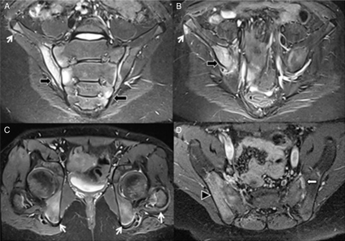
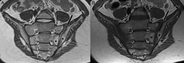
Spondyloarthropathy (SpA) is a group of inflammatory conditions that include spondylitis, sacroiliitis, asymmetrical peripheral arthritis and enthesitis. This condition is known as juvenile SpA when the diagnosis is made in patients up to 16 years of age. Enthesitis is a highly specific feature that occurs more often in juvenile SpA than in the adult form. In contrast to adult onset SpA, the initial manifestation of juvenile SpA rarely presents as inflammatory back pain. Peripheral arthritis is the more common presenting feature. We report a case of a 12-year-old boy who presented with a 1-year history of progressive low back pain, gluteal pain and thigh pain. There were no clinical symptoms of arthropathy of the distal extremities. MRI of the whole spine was performed twice, which, unfortunately, was unyielding. Finally, MRI of the sacroiliac joints revealed asymmetric sacroiliitis as well as enthesitis of the hips and pelvis. Further laboratory data showed negative rheumatoid factor and positive human leucocyte antigen (HLA) B27. A diagnosis of juvenile SpA with sacroiliitis and enthesitis was made. The imaging characteristics of juvenile SpA are highlighted.
2015 BMJ Publishing Group Ltd.
Figures
Similar articles
-
Persistent enthesitis and spondyloarthropathy: a case series of 71 Bangladeshi people.J Back Musculoskelet Rehabil. 2015;28(3):463-71. doi: 10.3233/BMR-140541. J Back Musculoskelet Rehabil. 2015. PMID: 25322736
-
Use of dynamic magnetic resonance imaging to detect sacroiliitis in HLA-B27 positive and negative children with juvenile arthritides.J Rheumatol. 1998 Mar;25(3):556-64. J Rheumatol. 1998. PMID: 9517781
-
Comparison of patients with familial Mediterranean fever accompanied with sacroiliitis and patients with juvenile spondyloarthropathy.Clin Exp Rheumatol. 2017 Nov-Dec;35 Suppl 108(6):124-127. Epub 2017 Sep 26. Clin Exp Rheumatol. 2017. PMID: 28980897
-
A critical overview of the imaging arm of the ASAS criteria for diagnosing axial spondyloarthritis: what the radiologist should know.Diagn Interv Radiol. 2012 Nov-Dec;18(6):555-65. doi: 10.4261/1305-3825.DIR.5732-12.0. Epub 2012 Apr 6. Diagn Interv Radiol. 2012. PMID: 22484991 Review.
-
Update on juvenile spondyloarthritis.Rheum Dis Clin North Am. 2013 Nov;39(4):767-88. doi: 10.1016/j.rdc.2013.06.002. Epub 2013 Aug 24. Rheum Dis Clin North Am. 2013. PMID: 24182854 Free PMC article. Review.
References
Publication types
MeSH terms
LinkOut - more resources
Full Text Sources
Other Literature Sources
Medical
Research Materials