

Congenital Giant Keratinous Cyst Mimicking Lipoma: Case Report and Review
- PMID: 26677303
- PMCID: PMC4681229
- DOI: 10.4103/0019-5154.169160
Congenital Giant Keratinous Cyst Mimicking Lipoma: Case Report and Review
Abstract
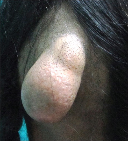
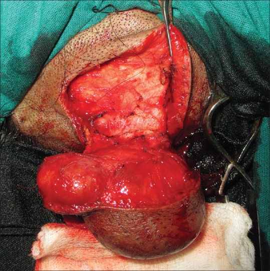
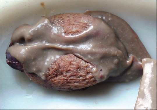
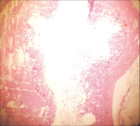
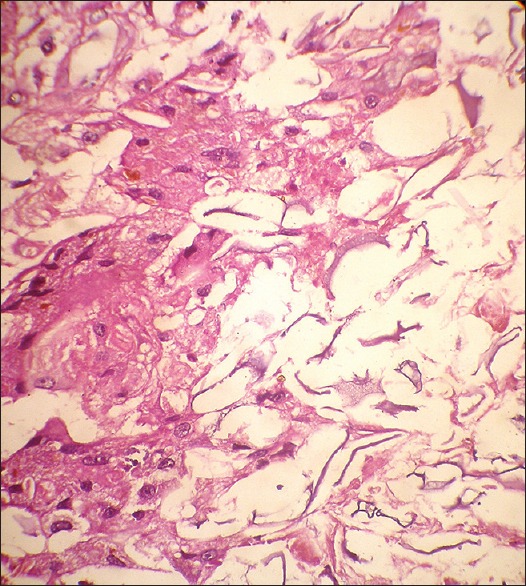
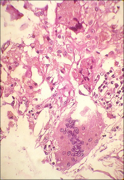
Epidermal cysts represent the most common cutaneous cysts. They arise following a localized inflammation of the hair follicle and occasionally after the implantation of the epithelium, following a trauma or surgery. Conventional epidermal cysts are about 5 cm in diameter; however, rare reports of cysts more than 5 cm are reported in the literature and are referred as "Giant epidermal cysts." Epidermal cysts although common, can mimic other common benign lesions in the head and neck area. A thorough clinico-pathologic investigation is needed to diagnose these cutaneous lesions as they differ in their biologic behavior, treatment, and prognosis. We report a case of a giant epidermoid cyst in the scalp area of a young female patient which mimicked lipoma on clinical, as well as cyotological examination. We also present a brief review of epidermal cysts, their histopathological differential diagnosis, and their malignant transformation.
Keywords: Giant epidermal cyst; histopathology; keratinous cyst; scalp.
Figures
Similar articles
-
A Rare Transformation of Epidermoid Cyst into Squamous Cell Carcinoma: A Case Report with Literature Review.Am J Case Rep. 2019 Aug 3;20:1141-1143. doi: 10.12659/AJCR.912828. Am J Case Rep. 2019. PMID: 31375657 Free PMC article. Review.
-
A Rare Case of an Epidermoid Cyst in the Parotid Gland - which was Diagnosed by Fine Needle Aspiration Cytology.J Clin Diagn Res. 2013 Mar;7(3):550-2. doi: 10.7860/JCDR/2013/4857.2822. Epub 2013 Mar 1. J Clin Diagn Res. 2013. PMID: 23634420 Free PMC article.
-
A Rare Presentation of a Giant Epidermoid Inclusion Cyst Mimicking Malignancy.J Foot Ankle Surg. 2018 Mar-Apr;57(2):421-426. doi: 10.1053/j.jfas.2017.09.005. Epub 2017 Dec 15. J Foot Ankle Surg. 2018. PMID: 29254851
-
Giant earlobe epidermoid cyst.J Cutan Aesthet Surg. 2012 Jan;5(1):38-9. doi: 10.4103/0974-2077.94342. J Cutan Aesthet Surg. 2012. PMID: 22557855 Free PMC article.
-
Epidermoid Cyst Arising on the Body of the Tongue: Case Report and Literature Review.J Nippon Med Sch. 2018;85(6):343-346. doi: 10.1272/jnms.JNMS.2018_85-56. J Nippon Med Sch. 2018. PMID: 30568062 Review.
Cited by
-
A Rare Transformation of Epidermoid Cyst into Squamous Cell Carcinoma: A Case Report with Literature Review.Am J Case Rep. 2019 Aug 3;20:1141-1143. doi: 10.12659/AJCR.912828. Am J Case Rep. 2019. PMID: 31375657 Free PMC article. Review.
-
A giant epidermal cyst preoperatively diagnosed by core needle biopsy.J Surg Case Rep. 2025 Feb 27;2025(2):rjaf088. doi: 10.1093/jscr/rjaf088. eCollection 2025 Feb. J Surg Case Rep. 2025. PMID: 40040767 Free PMC article.
-
An Unusual Presentation of Keratinous Cyst - A Case Report.J Orthop Case Rep. 2025 Aug;15(8):145-149. doi: 10.13107/jocr.2025.v15.i08.5918. J Orthop Case Rep. 2025. PMID: 40786793 Free PMC article.
-
Analysis of related factors between the occurrence of secondary epidermoid cyst of penis and circumcision.Sci Rep. 2022 Aug 9;12(1):13563. doi: 10.1038/s41598-022-16876-y. Sci Rep. 2022. PMID: 35945421 Free PMC article.
References
-
- Zuber TJ. Minimal excision technique for epidermoid (sebaceous) cysts. Am Fam Physician. 2002;65:1409. - PubMed
-
- Nicolaides N, Levan NE, Fu HC. The lipid pattern of the wen (keratinous cyst of the skin) J Invest Dermatol. 1968;50:189–94. - PubMed
-
- Mackie RM. Tumours of the skin. In: Rook A, Wilkinson DS, Ebling FJ, editors. Textbook of Dermatology. 4th ed. Vol. 3. St. Louis: Blackwell Mosby Book Distributors; 1986. pp. 2405–6.
-
- Huang CC, Ko SF, Huang HY, Ng SH, Lee TY, Lee YW, et al. Epidermal cysts in the superficial soft tissue: Sonographic features with an emphasis on the pseudotestis pattern. J Ultrasound Med. 2011;30:11–7. - PubMed
LinkOut - more resources
Full Text Sources
Other Literature Sources