

Evaluation of serum prolidase activity in patients with slow coronary flow
- PMID: 26677361
- PMCID: PMC4631735
- DOI: 10.5114/pwki.2015.54015
Evaluation of serum prolidase activity in patients with slow coronary flow
Abstract
Introduction: Slow coronary flow (SCF) is described as the slow passage of contrast to distal coronaries despite anatomically normal coronary arteries. It has been shown that increased serum prolidase activity (SPA) correlates with collagen turnover. Increased collagen turnover might be associated with the development of atherosclerotic plaques.
Aim: To investigate the relationship between serum prolidase activity and slow coronary flow.
Material and methods: This cross-sectional study included 40 SCF patients (mean age: 55.0 ±9.5 years, 20 females) and 40 controls (mean age: 53.9 ±8.2 years, 21 females) with normal coronary anatomy and normal coronary flow. The Thrombolysis in Myocardial Infarction (TIMI) frame-count (TFC) method was used for SCF diagnosis. Serum prolidase activity was measured spectrophotometrically, and the relevant parameters were compared between the groups.
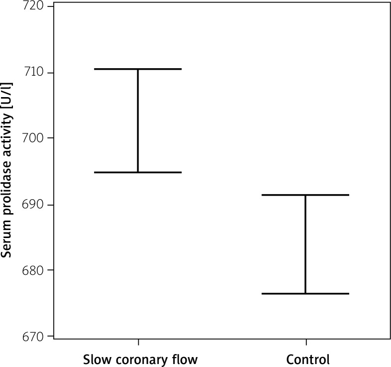
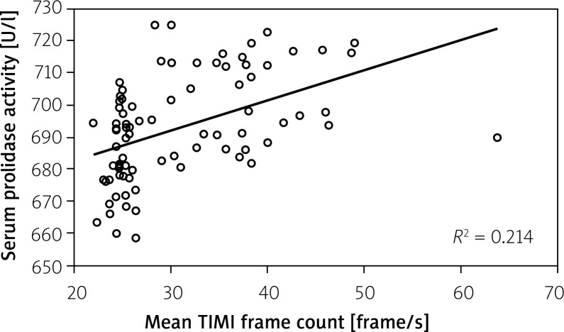
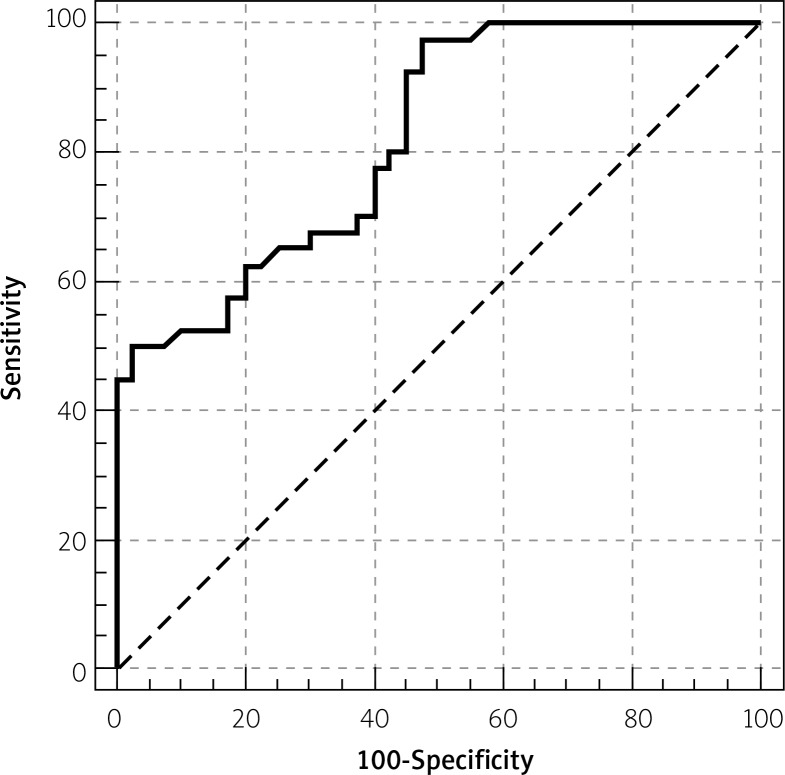
Results: There were no statistically significant differences between the SCF and control groups in terms of basic demographic, clinical, and laboratory data. However, the SPA was significantly higher in the SCF group compared to the control (702.7 ±13.8 and 683.9 ±13.2 respectively, p<0.001). Serum prolidase activity was significantly correlated with the mean TFC (r=0.463, p<0.001). The overall findings of this study support the predictive accuracy of the serum prolidase activity in our cohort, with a statistically significant ROC value of 681.3.
Conclusions: Our study showed that SPA was increased in SCF patients. The activity of this enzyme was significantly correlated with the mean TFC.
Keywords: collagen; serum prolidase activity; slow coronary flow.
Figures
Similar articles
-
Assessment of risk factors and left ventricular function in patients with slow coronary flow.Heart Vessels. 2016 Mar;31(3):288-97. doi: 10.1007/s00380-014-0606-4. Epub 2014 Dec 5. Heart Vessels. 2016. PMID: 25475386
-
Association of paraoxonase activity and coronary blood flow.Atherosclerosis. 2008 Mar;197(1):257-63. doi: 10.1016/j.atherosclerosis.2007.04.004. Epub 2007 May 30. Atherosclerosis. 2008. PMID: 17537444
-
Increased mean platelet volume in patients with slow coronary flow.Platelets. 2009 Feb;20(1):23-8. doi: 10.1080/09537100802458969. Platelets. 2009. PMID: 19172518
-
Relationship between elevated serum gamma-glutamyltransferase activity and slow coronary flow.Turk Kardiyol Dern Ars. 2009 Apr;37(3):168-73. Turk Kardiyol Dern Ars. 2009. PMID: 19553739
-
Interaction of plasma homocysteine and thyroid hormone concentrations in the pathogenesis of the slow coronary flow phenomenon.Cardiology. 2007;108(3):186-92. doi: 10.1159/000096687. Epub 2006 Nov 3. Cardiology. 2007. PMID: 17085937
Cited by
-
Circulating Prolidase Activity in Patients with Myocardial Infarction.Front Cardiovasc Med. 2017 Jul 31;4:50. doi: 10.3389/fcvm.2017.00050. eCollection 2017. Front Cardiovasc Med. 2017. PMID: 28824924 Free PMC article.
-
Association of serum prolidase activity in patients with isolated coronary artery ectasia.Anatol J Cardiol. 2018 Feb;19(2):110-116. doi: 10.14744/AnatolJCardiol.2017.8160. Epub 2018 Jan 17. Anatol J Cardiol. 2018. PMID: 29339675 Free PMC article.
-
Serum prolidase activity in patients with cardiac syndrome X.North Clin Istanb. 2020 Aug 17;7(5):471-477. doi: 10.14744/nci.2020.09086. eCollection 2020. North Clin Istanb. 2020. PMID: 33163883 Free PMC article.
References
-
- Tambe AA, Demany MA, Zimmerman HA, Mascarenhas E. Angina pectoris and slow flow velocity of dye in coronary arteries – a new angiographic finding. Am Heart J. 1972;84:66–71. - PubMed
-
- Kemp HG, Jr, Vokonas PS, Cohn PF, Gorlin R. The anginal syndrome associated with normal coronary arteriograms. Report of a six year experience. Am J Med. 1973;54:735–42. - PubMed
-
- Pekdemir H, Cin VG, Cicek D, et al. Slow coronary flow may be a sign of diffuse atherosclerosis. Contribution of FFR and IVUS. Acta Cardiol. 2004;59:127–33. - PubMed
-
- Mosseri M, Yarom R, Gotsman MS, Hasin Y. Histologic evidence for small-vessel coronary artery disease in patients with angina pectoris and patent large coronary arteries. Circulation. 1986;7:964–72. - PubMed
-
- Mangieri M, Macchiarelli G, Ciavolella M, et al. Slow coronary flow: clinical and histopathological features in patients with otherwise normal epicardial coronary arteries. Cathet Cardiovasc Diagn. 1996;37:375–81. - PubMed
LinkOut - more resources
Full Text Sources
Other Literature Sources