

The utility of cardiac magnetic resonance imaging in Kounis syndrome
- PMID: 26677363
- PMCID: PMC4631737
- DOI: 10.5114/pwki.2015.54017
The utility of cardiac magnetic resonance imaging in Kounis syndrome
Abstract
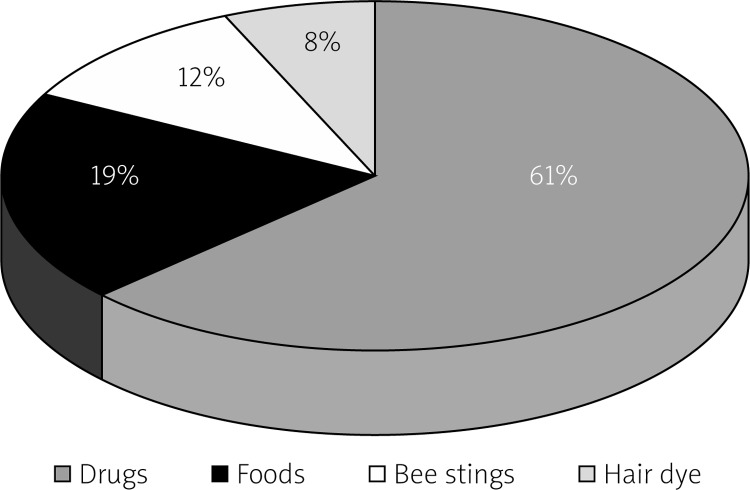
Introduction: Current diagnostic measurements used to assess myocardial involvement in Kounis syndrome, such as electrocardiography (ECG), cardiac enzymes, and troponin levels, are relatively insensitive to small but potentially significant functional change. According to our review of the literature, there has been no study using magnetic resonance imaging (MRI) on Kounis syndrome except for one case report.
Aim: To identify the findings of dynamic contrast-enhanced magnetic resonance imaging (CE-MRI) in patients with Kounis syndrome (KS) type 1.
Material and methods: We studied 26 patients (35 ±11.5 years, 53.8% male) with known or suspected KS type 1. The patients underwent precontrast, first-pass, and delayed enhancement cardiac MRI (DE-MRI). Contrast enhancement patterns, edema, hypokinesia, and localization for myocardial lesions were evaluated in all KS type 1 patients.
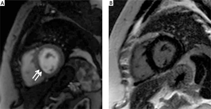
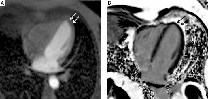
Results: Contrast-enhanced magnetic resonance imaging demonstrated an early-phase subendocardial contrast defect, and T2-weighted images showed high-signal intensity consistent with edema in lesion areas. None of the lesion areas was found upon contrast enhancement on DE-MRI. The area of early-phase subendocardial contrast defect was reported as follows: the interventricular septum in 14 (53.8%) patients, the left ventricular lateral wall in 8 (30.7%), and the left ventricular apex in 4 (15.4%).
Conclusions: Dynamic cardiac MR imaging is a reliable tool for assessing cardiac involvement in Kounis syndrome. Delayed contrast-enhanced images show normal washout in the subendocardial lesion area in patients with Kounis syndrome type 1.
Keywords: Kounis syndrome; cardiac magnetic resonance imaging; hypersensitivity.
Figures
Similar articles
-
Two questions for Kounis syndrome: can we use magnetic resonance imaging in the diagnosis and does ST elevation correlates with troponin levels?Am J Emerg Med. 2012 Nov;30(9):2086.e5-7. doi: 10.1016/j.ajem.2011.12.016. Epub 2012 Mar 3. Am J Emerg Med. 2012. PMID: 22386343
-
A Curious Case of Coronary Vasospasm with Cardiogenic Shock: Type 1 Kounis Syndrome Complicated by Eosinophilic Myocarditis.Cureus. 2019 Apr 22;11(4):e4522. doi: 10.7759/cureus.4522. Cureus. 2019. PMID: 31259131 Free PMC article.
-
Acute myocarditis or the kounis syndrome: role of cardiac MRI and speckle-tracking echocardiography in diagnosis.Res Cardiovasc Med. 2014 Nov 26;3(4):e25715. doi: 10.5812/cardiovascmed.25715. eCollection 2014 Nov. Res Cardiovasc Med. 2014. PMID: 25780783 Free PMC article.
-
[Optimal use of contrast medium in contrast enhanced MR imaging of the heart].Nihon Igaku Hoshasen Gakkai Zasshi. 2002 Oct;62(12):682-9. Nihon Igaku Hoshasen Gakkai Zasshi. 2002. PMID: 12462069 Review. Japanese.
-
[Patterns of delayed-enhancement in MRI of ischemic and non-ischemic cardiomyopathies].Rofo. 2007 Jan;179(1):21-30. doi: 10.1055/s-2006-927204. Rofo. 2007. PMID: 17203440 Review. German.
Cited by
-
Case Report: Perioperative Kounis Syndrome in an Adolescent With Congenital Glaucoma.Front Cardiovasc Med. 2021 Sep 10;8:676188. doi: 10.3389/fcvm.2021.676188. eCollection 2021. Front Cardiovasc Med. 2021. PMID: 34568441 Free PMC article.
-
Recurrent Kounis Syndrome: A Case Report and Literature Review.J Clin Med. 2024 Mar 13;13(6):1647. doi: 10.3390/jcm13061647. J Clin Med. 2024. PMID: 38541873 Free PMC article. Review.
-
Contrast Media Induced Kounis Syndrome: A Case Report.Diagnostics (Basel). 2019 Oct 18;9(4):154. doi: 10.3390/diagnostics9040154. Diagnostics (Basel). 2019. PMID: 31635242 Free PMC article.
-
Kounis syndrome: ST elevations in the setting of anaphylaxis.J Allergy Clin Immunol Glob. 2023 Jul 24;2(4):100152. doi: 10.1016/j.jacig.2023.100152. eCollection 2023 Nov. J Allergy Clin Immunol Glob. 2023. PMID: 37781662 Free PMC article.
-
Kounis Syndrome: A Case Report and a Review of Recent Literature.Cureus. 2024 Jul 16;16(7):e64627. doi: 10.7759/cureus.64627. eCollection 2024 Jul. Cureus. 2024. PMID: 39149660 Free PMC article.
References
-
- Kounis NG. Cardiovascular disease and allergy: angina pectoris myocardial infarction. In: Hamilton K, Simpson K, editors. The experts speak: the role of nutrition in medicine. Sacramento, Calif: IT Services; 1997. p. 36.
-
- Lopez PR, Peiris AN. Kounis syndrome. South Med J. 2010;103:1148–55. - PubMed
-
- Biteker M. Current understanding of Kounis syndrome. Expert Rev Clin Immunol. 2010;6:777–88. - PubMed
-
- Biteker M. A new classification of Kounis syndrome. Int J Cardiol. 2010;145:553. - PubMed
-
- Park JM, Cho J, Chung SP, Kim MJ. Kounis syndrome captured by coronary angiography computed tomography. Am J Emerg Med. 2010;28:640.e5–8. - PubMed
LinkOut - more resources
Full Text Sources
Other Literature Sources
Miscellaneous