

Abnormal wiring of the connectome in adults with high-functioning autism spectrum disorder
- PMID: 26677408
- PMCID: PMC4681075
- DOI: 10.1186/s13229-015-0058-4
Abnormal wiring of the connectome in adults with high-functioning autism spectrum disorder
Abstract
Background: Recent brain imaging findings suggest that there are widely distributed abnormalities affecting the brain connectivity in individuals with autism spectrum disorder (ASD). Using graph theoretical analysis, it is possible to investigate both global and local properties of brain's wiring diagram, i.e., the connectome.
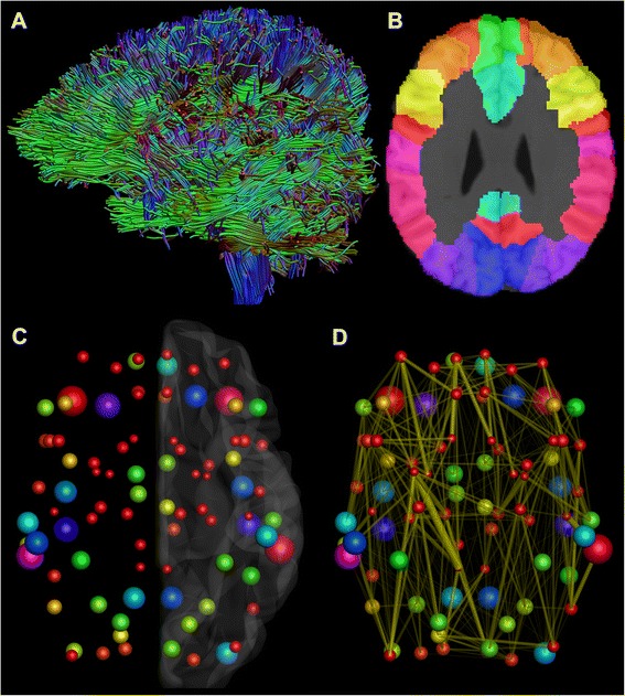
Methods: We acquired diffusion-weighted magnetic resonance imaging data from 14 adult males with high-functioning ASD and 19 age-, gender-, and IQ-matched controls. As with diffusion tensor imaging-based tractography, it is not possible to detect complex (e.g., crossing) fiber configurations, present in 60-90 % of white matter voxels; we performed constrained spherical deconvolution-based whole brain tractography. Unweighted and weighted structural brain networks were then reconstructed from these tractography data and analyzed with graph theoretical measures.
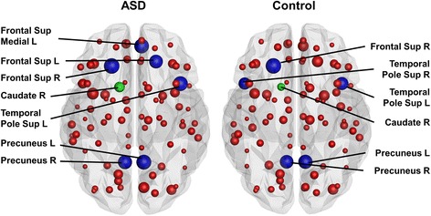
Results: In subjects with ASD, global efficiency was significantly decreased both in the unweighted and the weighted networks, normalized characteristic path length was significantly increased in the unweighted networks, and strength was significantly decreased in the weighted networks. In the local analyses, betweenness centrality of the right caudate was significantly increased in the weighted networks, and the strength of the right superior temporal pole was significantly decreased in the unweighted networks in subjects with ASD.
Conclusions: Our findings provide new insights into understanding ASD by showing that the integration of structural brain networks is decreased and that there are abnormalities in the connectivity of the right caudate and right superior temporal pole in subjects with ASD.
Keywords: Autism spectrum disorder; Brain networks; Connectivity; Connectome; Diffusion magnetic resonance imaging; Graph theoretical analysis; Tractography; White matter tract.
Figures

Similar articles
-
Reproducibility and intercorrelation of graph theoretical measures in structural brain connectivity networks.Med Image Anal. 2019 Feb;52:56-67. doi: 10.1016/j.media.2018.10.009. Epub 2018 Oct 26. Med Image Anal. 2019. PMID: 30471463
-
Constrained spherical deconvolution-based tractography and tract-based spatial statistics show abnormal microstructural organization in Asperger syndrome.Mol Autism. 2015 Jan 16;6:4. doi: 10.1186/2040-2392-6-4. eCollection 2015. Mol Autism. 2015. PMID: 25874076 Free PMC article.
-
White matter and visuospatial processing in autism: a constrained spherical deconvolution tractography study.Autism Res. 2013 Oct;6(5):307-19. doi: 10.1002/aur.1290. Epub 2013 Mar 18. Autism Res. 2013. PMID: 23509018
-
The functional brain connectome of the child and autism spectrum disorders.Acta Paediatr. 2016 Sep;105(9):1024-35. doi: 10.1111/apa.13484. Epub 2016 Jun 23. Acta Paediatr. 2016. PMID: 27228241 Review.
-
Altered white matter connectivity as a neural substrate for social impairment in Autism Spectrum Disorder.Cortex. 2015 Jan;62:158-81. doi: 10.1016/j.cortex.2014.10.014. Epub 2014 Nov 5. Cortex. 2015. PMID: 25433958 Review.
Cited by
-
Using tissue microstructure and multimodal MRI to parse the phenotypic heterogeneity and cellular basis of autism spectrum disorder.J Child Psychol Psychiatry. 2022 Aug;63(8):855-870. doi: 10.1111/jcpp.13531. Epub 2021 Nov 11. J Child Psychol Psychiatry. 2022. PMID: 34762311 Free PMC article.
-
Altered brain network organization in adults with Asperger's syndrome: decreased connectome transitivity and assortativity with increased global efficiency.Front Psychiatry. 2023 Aug 17;14:1223147. doi: 10.3389/fpsyt.2023.1223147. eCollection 2023. Front Psychiatry. 2023. PMID: 37701094 Free PMC article.
-
Shared and Distinct Topologically Structural Connectivity Patterns in Autism Spectrum Disorder and Attention-Deficit/Hyperactivity Disorder.Front Neurosci. 2021 Jun 11;15:664363. doi: 10.3389/fnins.2021.664363. eCollection 2021. Front Neurosci. 2021. PMID: 34177449 Free PMC article.
-
Pediatric Traumatic Brain Injury and Autism: Elucidating Shared Mechanisms.Behav Neurol. 2016;2016:8781725. doi: 10.1155/2016/8781725. Epub 2016 Dec 15. Behav Neurol. 2016. PMID: 28074078 Free PMC article. Review.
-
Gender differences in the structural connectome of the teenage brain revealed by generalized q-sampling MRI.Neuroimage Clin. 2017 May 22;15:376-382. doi: 10.1016/j.nicl.2017.05.014. eCollection 2017. Neuroimage Clin. 2017. PMID: 28580294 Free PMC article.
References
Grants and funding
LinkOut - more resources
Full Text Sources
Other Literature Sources
Research Materials
