. 2015 Dec 8;5(6):996-1009.
doi: 10.1016/j.stemcr.2015.10.012. Epub 2015 Nov 19.

Reduced Adult Hippocampal Neurogenesis and Cognitive Impairments following Prenatal Treatment of the Antiepileptic Drug Valproic Acid

Affiliations

Reduced Adult Hippocampal Neurogenesis and Cognitive Impairments following Prenatal Treatment of the Antiepileptic Drug Valproic Acid

Berry Juliandi et al. Stem Cell Reports. .

Abstract

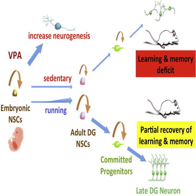
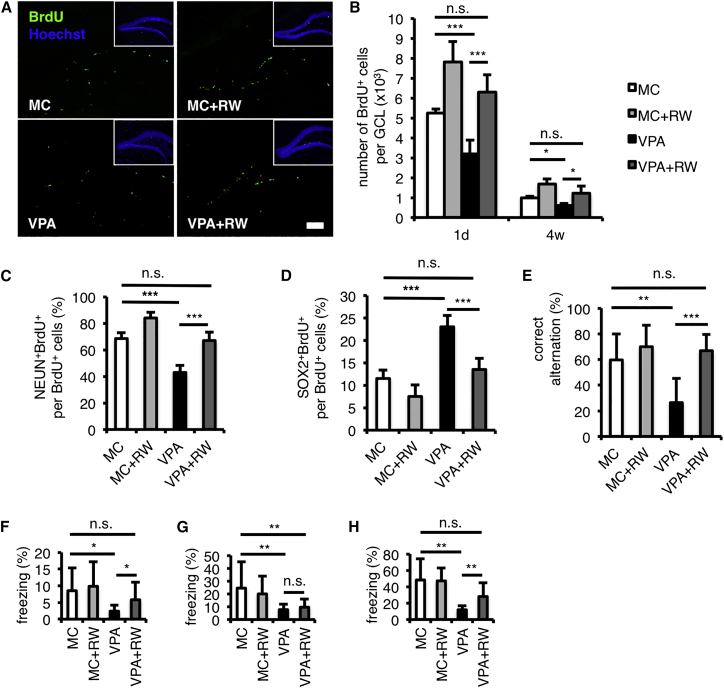
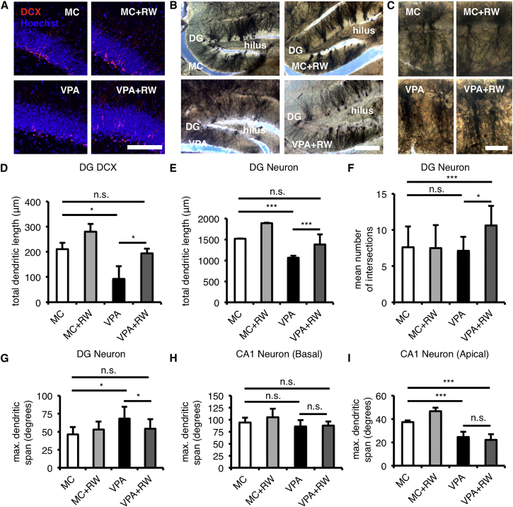
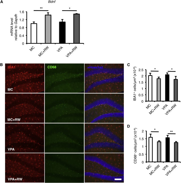
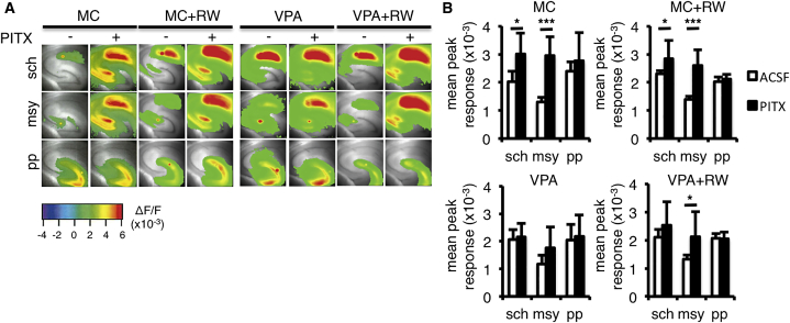
Prenatal exposure to valproic acid (VPA), an established antiepileptic drug, has been reported to impair postnatal cognitive function in children born to VPA-treated epileptic mothers. However, how these defects arise and how they can be overcome remain unknown. Using mice, we found that comparable postnatal cognitive functional impairment is very likely correlated to the untimely enhancement of embryonic neurogenesis, which led to depletion of the neural precursor cell pool and consequently a decreased level of adult neurogenesis in the hippocampus. Moreover, hippocampal neurons in the offspring of VPA-treated mice showed abnormal morphology and activity. Surprisingly, these impairments could be ameliorated by voluntary running. Our study suggests that although prenatal exposure to antiepileptic drugs such as VPA may have detrimental effects that persist until adulthood, these effects may be offset by a simple physical activity such as running.


Figures

None
Graphical abstract
Figure 1
Figure 1
Prenatal VPA Treatment Enhances Untimely Embryonic Neurogenesis (A) Experimental timeline of prenatal VPA treatment. E, embryonic day. (B) The region highlighted by the dashed black rectangle in E15.5 forebrain sections was used for analysis in (C) and (D). The image is modified from the Electronic Prenatal Mouse Brain Atlas. Scale bar, 1 mm. (C) The thickness of the region stained by immature neuron marker β-tubulin isotype III (TUJ1; magenta) is increased in the cortical hem of VPA-treated mice. Scale bar, 100 μm. (D) VPA treatment increases the production of TBR2+ intermediate neuronal progenitors (red) born in the dentate neuroepithelium. Scale bar, 100 μm. (E) Quantification of the density of TBR2+ in (D). (F–I) The proportion of E14.5 BrdU-labeled NPCs (red) expressing NPC marker SOX2 (green; F and H) is reduced, while the ones that had differentiated into NEUN+ neurons are increased at P7 hippocampus after embryonic VPA treatment (green; G and I). Scale bars, 100 μm. (J and K) Embryonic VPA treatment reduces the number of highly proliferating NPCs (red) labeled by 30 min single-pulse BrdU injection in the P7 DG. See also Figure S1 for other immunostaining data in the cortex of embryonic forebrain. MC, prenatal methylcellulose (vehicle); VPA, prenatal valproic acid. Data are represented as means. n = 3 for each group. Error bars indicate the SD. p < 0.05, ∗∗p < 0.01, ∗∗∗p < 0.001, two-tailed t test. Scale bar, 50 μm. See also Figure S1.
Figure 2
Figure 2
VPA-Treated Mice Perform Poorly in Learning and Memory Tests (A) Experimental timeline of prenatal VPA treatment and postnatal behavior tests. E, embryonic day; w, weeks old. (B) VPA-treated mice have a lower correct-arm alternation than MC-treated mice (control). (C–E) VPA-treated mice have a lower freezing response than MC-treated mice (control) in conditioning (C; day 1), contextual (D; day 2), and cued fear associative tests (E; day 3). See also Figures S4A, S4C, and S4E for time course of freezing response and Table S1 for a summary of behavior data. MC, prenatal methylcellulose (vehicle); VPA, prenatal valproic acid. Data are represented as means. n = 12 for each group. Error bars indicate the SD. p < 0.05, ∗∗p < 0.01, two-tailed t test. See also Figure S4 and Table S1.
Figure 3
Figure 3
Prenatal VPA Treatment Has the Long-Term Effect on the Adult Neurogenesis (A) Experimental timeline of prenatal VPA treatment and adult neurogenesis analysis. E, embryonic day; P, postnatal day; 1d, 1 day after the last intraperitoneal (i.p.) BrdU injection; 4w, 4 weeks after the last i.p. BrdU injection. (B) Representative images of brain sections including the hippocampal DG stained for BrdU (red) and with Hoechst 33258 (blue),1 day after the last BrdU injection. See also Figure S2A for KI-67 staining. (C) Quantification of BrdU+ cells in the granule cell layer (GCL), 1 day (1d; n = 8 for each group) and 4 weeks (4w; n = 8 for each group) after the last BrdU injection, shows a reduction of BrdU+ cells in the hippocampus. See also Figure S2B for quantification of KI-67+ cells in the GCL. (D) NPC differentiation into NEUN+ neurons and S100β+ astrocytes is impaired in VPA-treated mice, as shown by a reduced proportion of marker-positive and BrdU+ cells among total BrdU+ cells at 4 weeks after the last BrdU injection (n = 4 for each group). (E) BrdU+ cell survival is similar in VPA- and MC-treated mice. Quantification of BrdU+ cell survival in each group as a percentage of BrdU+ cells at 4w relative to BrdU+ cells at 1d (n = 8 for each group). MC, prenatal methylcellulose (vehicle); VPA, prenatal valproic acid. Data are represented as means. Error bars indicate the SD. p < 0.05, ∗∗p < 0.01, ∗∗∗p < 0.001, n.s., not significantly different, two-tailed t test. Scale bar, 100 μm. See also Figures S2 and S3.
Figure 4
Figure 4
Voluntary Running Restores Adult Neurogenesis and Cognitive Deficiencies of VPA-Treated Mice (A) Representative images of brain sections including the hippocampal DG stained for BrdU (green) and with Hoechst 33258 (blue), 1 day after the last BrdU injection. See Figure 2A for the experimental timeline. (B) Quantification of BrdU+ in the granule cell layer (GCL), 1 day (1d; n = 8 for each group) and 4 weeks (4w; n = 8 for each group) after the last BrdU injection, shows an increased number of BrdU+ cells in the hippocampus after voluntary running. (C and D) Voluntary running recovers the proportion of BrdU+ cells that had differentiated into NEUN+ neurons (C) and the ones that still expressed SOX2 (D) among total BrdU+ cells at 4 weeks after the last BrdU injection (n = 4 for each group). (E) Reduction of correct-arm alternation in Y-maze tests of VPA-treated mice is recovered by voluntary running (n = 7 for MC and VPA + RW; n = 8 for MC + RW and VPA). (F–H) Voluntary running recovers the freezing response in conditioning (F; day 1) and in cued fear associative tests (H; day 3), but not in contextual fear associative tests (G; day 2; n = 7 for MC and VPA + RW; n = 8 for MC + RW and VPA). See also Figures S4B, S4D, and S4F for the time course of the freezing response and Table S2 for a summary of behavior data. MC, prenatal methylcellulose (vehicle); MC + RW, prenatal methylcellulose and postnatal running; VPA, prenatal valproic acid; VPA + RW, prenatal valproic acid and postnatal running. Data are represented as means. Error bars indicate the SD. p < 0.05, ∗∗p < 0.01, ∗∗∗p < 0.001, n.s., not significantly different, two-tailed t test. Scale bar, 100 μm. See also Figure S4 and Table S2.
Figure 5
Figure 5
Voluntary Running Restores Neuronal Morphology in VPA-Treated Mice (A) Impaired morphology of DCX+ young neurons in the DG of VPA-treated mice is recovered by voluntary running (n = 4 for each group). Scale bar, 100 μm. (B) Impaired morphology of Golgi-Cox stained neurons in the DG of VPA-treated mice is recovered by voluntary running (n = 3 for each group). Note that voluntary running recovered non-molecular layer-oriented dendrites in VPA-treated mice to molecular layer-oriented ones. Scale bar, 100 μm. (C) Impaired morphology of Golgi-Cox stained neurons in the CA1 of VPA-treated mice is not recovered by voluntary running (n = 3 for each group). Note that the less-ramified and straighter apical dendrites in VPA-treated mice could not be recovered by voluntary running. Scale bar, 50 μm. (D–F) Voluntary running recovers total dendritic length of DCX+ young neurons (D) and Golgi-Cox stained neurons (E) and increases dendritic complexity of Golgi-Cox stained neurons (F) in the DG of VPA-treated mice. See also Figures S5A and S5B for Sholl analysis. (G–I) Abnormal dendritic span of DG neurons (G), but not of apical dendritic span of CA1 neurons (I), is recovered by voluntary running in VPA-treated mice, while basal dendrites of CA1 neurons show similar dendritic span across groups (H). MC, prenatal methylcellulose (vehicle); MC + RW, prenatal methylcellulose and postnatal running; VPA, prenatal valproic acid; VPA + RW, prenatal valproic acid and postnatal running. Data are represented as means. Error bars indicate the SD. p < 0.05, ∗∗∗p < 0.001, n.s., not significantly different, two-tailed t test. See also Figure S5.
Figure 6
Figure 6
Voluntary Running Increases the Bdnf Expression Level and Reduces Microglia and Activated Microglia in the Hippocampus (A) The expression level of brain-derived neurotrophic factor (Bdnf) was increased by voluntary running in both MC- and VPA-treated mice. (B–D) Voluntary running reduced the number of IBA1+ microglia (red; B and C) and CD68+-activated microglia (green; B and D) in both MC- and VPA-treated mice. MC, prenatal methylcellulose (vehicle); MC + RW, prenatal methylcellulose and postnatal running; VPA, prenatal valproic acid; VPA + RW, prenatal valproic acid and postnatal running; Gapdh, glyceraldehyde 3-phosphate dehydrogenase. Data are represented as means. n = 3 for each group. Error bars indicate the SD. p < 0.05, ∗∗p < 0.01, two-tailed t test. Scale bar, 100 μm.
Figure 7
Figure 7
Voluntary Running Restores Neuronal Activity in VPA-Treated Mice (A) Representative pseudocolor activity map images of brain slices including the hippocampus show that voluntary running can only recover the impairment of GABAA receptor-mediated inhibition in the mossy fiber pathway (msy) of VPA-treated mice, after treatment with the GABAA receptor channel antagonist picrotoxin (PITX) (n = 6 for MC, n = 9 for MC + RW, n = 7 for VPA, n = 8 for VPA + RW). Electrical stimulation was applied to Schaffer collateral afferents at the CA3/CA1 border of CA1 (sch); to the granule cell layer to stimulate the mossy fiber pathway (msy); and to the molecular layer of the upper blade in the DG (pp). (B) Quantification of the neural response in artificial cerebrospinal fluid (ACSF), with (black bars) or without PITX (white bars; n = 6 for MC, n = 9 for MC + RW, n = 7 for VPA, n = 8 for VPA + RW). Note that although the augmentation of the neural response caused by GABAA receptor-mediated inhibition with PITX application seen in sch and msy was abolished in VPA-treated mice, voluntary running could restore the augmentation only in the msy. MC, prenatal methylcellulose (vehicle); MC + RW, prenatal methylcellulose and postnatal running; VPA, prenatal valproic acid; VPA + RW, prenatal valproic acid and postnatal running. Data are represented as means. Error bars indicate the SD. p < 0.05, ∗∗∗p < 0.001, two-tailed t test.

References

    1. Abematsu M., Tsujimura K., Yamano M., Saito M., Kohno K., Kohyama J., Namihira M., Komiya S., Nakashima K. Neurons derived from transplanted neural stem cells restore disrupted neuronal circuitry in a mouse model of spinal cord injury. J. Clin. Invest. 2010;120:3255–3266. - PMC - PubMed
    1. Alvarez-Buylla A., Lim D.A. For the long run: maintaining germinal niches in the adult brain. Neuron. 2004;41:683–686. - PubMed
    1. Balasubramaniyan V., Boddeke E., Bakels R., Küst B., Kooistra S., Veneman A., Copray S. Effects of histone deacetylation inhibition on neuronal differentiation of embryonic mouse neural stem cells. Neuroscience. 2006;143:939–951. - PubMed
    1. Battino D., Tomson T. Management of epilepsy during pregnancy. Drugs. 2007;67:2727–2746. - PubMed
    1. Bekinschtein P., Oomen C.A., Saksida L.M., Bussey T.J. Effects of environmental enrichment and voluntary exercise on neurogenesis, learning and memory, and pattern separation: BDNF as a critical variable? Semin. Cell Dev. Biol. 2011;22:536–542. - PubMed

Publication types

MeSH terms