Review
. 2016 Jan;41(1):94-105.
doi: 10.1016/j.tibs.2015.11.004. Epub 2015 Dec 9.

How Dynein Moves Along Microtubules

Affiliations
Review

How Dynein Moves Along Microtubules

Gira Bhabha et al. Trends Biochem Sci. 2016 Jan.

Abstract

Cytoplasmic dynein, a member of the AAA (ATPases Associated with diverse cellular Activities) family of proteins, drives the processive movement of numerous intracellular cargos towards the minus end of microtubules. Here, we summarize the structural and motile properties of dynein and highlight features that distinguish this motor from kinesin-1 and myosin V, two well-studied transport motors. Integrating information from recent crystal and cryoelectron microscopy structures, as well as high-resolution single-molecule studies, we also discuss models for how dynein biases its movement in one direction along a microtubule track, and present a movie that illustrates these principles.


Figures

Figure 1
Figure 1
Architecture of the dynein motor domain and the linker conformational change. Two crystal structures are shown: (Left panel) human cytoplasmic dynein 2 with ADP-vanadate, which is though to mimic ATP or ADP-Pi in AAA1 (PDB code: 4RH7) and (right panel) Dictyostelium cytoplasmic dynein with ADP bound in AAA1 (PDB code: 3VKH). AAA1 is the main hydrolytic site that drives the motility cycle. Transitions of the linker (magenta) between bent and straight conformations may be involved in driving dynein motility (see Figure 2). This figure was generated using ePMV [68].
Figure 2
Figure 2
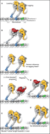
A model for dynein stepping along a microtubule. See also Movie S1. A) The sequence starts with a cytoplasmic dynein dimer with ADP bound in both motor domains (yellow) and both microtubule-binding domains (MTBDs) tightly bound to the microtubule, in the strong-binding state (indicated by “s” and darker blue color). One tubulin dimer in the microtubule is highlighted in green to allow easier identification of dynein progression towards the minus end. B) When ADP is exchanged for ATP in the leading head (red), conformational changes occur in the ring, which are communicated to the MTBD via a change in registry of the coiled-coil stalk, thus releasing the MTBD from the microtubule. This sequence triggers the reposition of the N terminus of the linker over AAA2/3 and a resultant rotation of the ring/MTBD and a slight forward re-positioning of the MTBD. The weakly bound motor domain (indicated by “w” and lighter blue color) is also subject to Brownian motion, which causes a two-dimensional search of the MTBD on top of the microtubule lattice. The weak-to-strong binding transition is favored with the stalk pointing backwards, and this locks a forward step in place by the front head (see also Figure 3). This asymmetry in the binding properties of the MTBD provides an additional proof-reading mechanism that favors a forward versus a backward step. C) This strong rebinding triggers the movement of the linker from AAA2/3 to AAA4/5. This minus-end directed shift of the N terminus of the linker reduces the slack in the connection between the motor domains and applies tension to the lagging motor domain. At this point, two potential subsequent steps are shown (D,E and D',E'). D and E) The rear head now detaches after ADP/ATP exchange and undergoes the same sequence of events described for the front head, again resulting in a step forward towards the front head and relieving the tension between the two motor domains. D’ and E’) The leading motor domain undergoes ADP/ATP exchange and detaches, but tension between the motor domains overrides the forward biasing mechanisms and the leading MTBD steps backward. In this instance, the homodimer does not undergo a net displacement from A to E’.
Figure 3
Figure 3
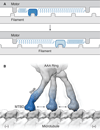
Asymmetry in polymer binding can create a directional bias. A) A depiction of a model by Andrew Huxley for muscle contraction [69]. In this model, which was developed before the myosin structure was known, the motor contains a small actin-binding element linked to a core by a spring-like element, which moves back-and-forth by thermal motion relative to the actin filament. The binding of this fluctuating element to the actin filament increases when displaced by thermal fluctuations in one direction (top panel, dark blue reveals a state with a high association rate). Conversely, its association rate is lower (bottom panel, light blue) when stretched in the opposite direction. Binding to the filament occurs spontaneously, but ATP energy is needed to dissociate the actomyosin bond, thus creating a mechanism that drives multiple force-generating cycles. B) A model for the binding of the MTBD to the microtubule. The stalk/MTBD can explore different binding sites by Brownian motion; the weak-to-strong transition of the MTBD (dark blue) is enhanced when displaced towards the minus end (left in this figure) but less likely when interacting with other binding sites (lighter blue).

References

    1. Allan VJ. Cytoplasmic dynein. Biochem. Soc. Trans. 2011;39:1169–1178. - PubMed
    1. Verhey KJ, et al. Kinesin assembly and movement in cells. Annu. Rev. Biophys. 2011;40:267–288. - PubMed
    1. Block SM. Kinesin motor mechanics: binding, stepping, tracking, gating, and limping. Biophys. J. 2007;92:2986–2995. - PMC - PubMed
    1. Vale RD, Milligan RA. The way things move: looking under the hood of molecular motor proteins. Science. 2000;288:88–95. - PubMed
    1. Hammer JA, Sellers JR. Walking to work: roles for class V myosins as cargo transporters. Nat. Rev. Mol. Cell Biol. 2012;13:13–26. - PubMed

Publication types