. 2015 Dec 17:13:107.
doi: 10.1186/s12915-015-0216-3.

A rapid and affordable screening platform for membrane protein trafficking

Affiliations

A rapid and affordable screening platform for membrane protein trafficking

Joshua C Snyder et al. BMC Biol. .

Abstract

Background: Membrane proteins regulate a diversity of physiological processes and are the most successful class of targets in drug discovery. However, the number of targets adequately explored in chemical space and the limited resources available for screening are significant problems shared by drug-discovery centers and small laboratories. Therefore, a low-cost and universally applicable screen for membrane protein trafficking was developed.

Results: This high-throughput screen (HTS), termed IRFAP-HTS, utilizes the recently described MarsCy1-fluorogen activating protein and the near-infrared and membrane impermeant fluorogen SCi1. The cell surface expression of MarsCy1 epitope-tagged receptors can be visualized by simple addition of SCi1. User-friendly, rapid, and quantitative detection occurs on a standard infrared western-blotting scanner. The reliability and robustness of IRFAP-HTS was validated by confirming human vasopressin-2 receptor and dopamine receptor-2 trafficking in response to agonist or antagonist. The IRFAP-HTS screen was deployed against the leucine-rich G protein-coupled receptor-5 (Lgr5). Lgr5 is expressed in stem cells, modulates Wnt/ß-catenin signaling, and is therefore a promising drug target. However, small molecule modulators have yet to be reported. The constitutive internalization of Lgr5 appears to be one primary mode through which its function is regulated. Therefore, IRFAP-HTS was utilized to screen 11,258 FDA-approved and drug-like small molecules for those that antagonize Lgr5 internalization. Glucocorticoids were found to potently increase Lgr5 expression at the plasma membrane.

Conclusion: The IRFAP-HTS platform provides a versatile solution for screening more targets with fewer resources. Using only a standard western-blotting scanner, we were able to screen 5,000 compounds per hour in a robust and quantitative assay. Multi-purposing standardly available laboratory equipment eliminates the need for idiosyncratic and more expensive high-content imaging systems. The modular and user-friendly IRFAP-HTS is a significant departure from current screening platforms. Small laboratories will have unprecedented access to a robust and reliable screening platform and will no longer be limited by the esoteric nature of assay development, data acquisition, and post-screening analysis. The discovery of glucocorticoids as modulators for Lgr5 trafficking confirms that IRFAP-HTS can accelerate drug-discovery and drug-repurposing for even the most obscure targets.


Figures
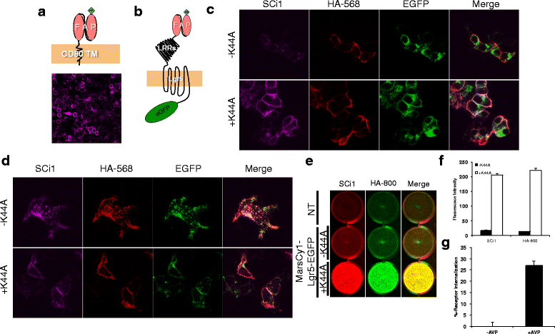
Fig. 1
Fig. 1
Quantitative scanning of MarsCy1-tagged membrane proteins in a modular plate-based format. a Top: Cartoon depicting HA-FAP fused to the N-terminus of the CD80 transmembrane domain. Bottom: Confocal imaging of FAP-CD80 transfected cells stained with SCi1. b Cartoon depicting HA-FAP fused to the N-terminus of Lgr5 and EGFP fused to the C-terminus. c Confocal imaging of FAP-tagged Lgr5-EGFP transfected cells that were co-labeled with SCi1 (magenta) and a primary HA-epitope antibody (Red, secondary retrieval – 568 nm) at 4 °C to block receptor internalization and were chased in (d) for 30 minutes at 37 °C to allow constitutive internalization of Lgr5 (EGFP, green) (K44A, A dominant-negative Dynamin I mutant that inhibits receptor endocytosis when over-expressed). e Infrared plate imaging of a 12-well plate (see Additional file 4: Figure S4 for entire plate and time-course) with FAP-tagged Lgr5 pulsed with SCi1 and a primary HA-epitope antibody at 4 °C and then fixed. Non-permeabilized cells were scanned on the plate at 700 nm (red, SCi1) and 800 nm (green, HA secondary retrieval-800 nm) (NT: non-transfected). f Integrated fluorescence intensity from (e). g MarsCy1-tagged hV2R was transiently transfected in HEK cells on a 24-well plate and stimulated with vehicle (–AVP) or the V2R ligand AVP [10 μM] for 1 hour. Cells were SCi1 stained, scanned, and quantified
Fig. 2
Fig. 2
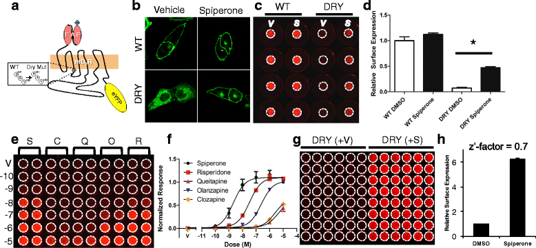
Monitoring cell surface rescue of a mutant intracellularly mis-localized membrane protein. a Cartoon depicting HA-FAP fused to the N-terminus of wild-type (WT) D2R or DRY-AAY (DRY) D2R and EYFP on the C-terminus. b Confocal imaging for EYFP-tagged WT-D2R or DRY-D2R treated with vehicle (DMSO) or the D2R antagonist spiperone [10 μM]. c Infrared plate imaging for membrane impermeable SCi1 bound to FAP-tagged WT- or DRY-D2R treated with DMSO or spiperone (S, 10 μM) overnight. d Quantification of triplicate experiments represented in panel (c) and normalized relative to WT-D2R surface expression (* denotes significant difference by ANOVA and post hoc Tukey-analysis). e Small-scale dose-response screen of overnight treatment with small molecule antagonists using quantitative infrared plate imaging (V, DMSO Vehicle; S, Spiperone; R, Risperidone; Q, Quetiapine; O, Olanzapine; C, Clozapine. 10-10 (-10) M to 10-5 (-5) M). f Quantification of triplicate experiments of the representative image in (e) and normalized to spiperone (S). g Infrared image of 10 μM spiperone DRY-D2R surface expression rescue for Z’-factor analysis. h Quantification of panel (g) and calculation of a Z’-factor (* denotes significant difference by student’s unpaired t-test)
Fig. 3
Fig. 3
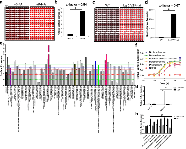
Screening for small molecule modulators of Lgr5 surface expression. a Infrared image of a stable U2OS cell line expressing MarsCy1-Lgr5 in a 384-well plate without (–K44A) and with transient transfection (+K44A) of Dynamin I K44A (NT, parental U2OS cells). b Quantification of (a) and Z’-factor analysis. c Infrared image of a stable U2OS cell lines expressing MarsCy1-Lgr5 compared to MarsCy1-Lgr5/V2R-tail (NT, parental U2OS cells). d Quantification of (c) and Z’-factor analysis. e A total of 91 hits were cherry picked and incubated overnight at 37 °C on stable MarsCy1-Lgr5-EGFP cells. Black bar, wild-type Lgr5; Hatched bar, +K44A control; Pink bar, autofluorescent compounds (violet, yellow, blue, and green bars as in 3f). Hits were measured against the ± 1, 2, 3 standard deviations from wild-type DMSO mean (green, blue, and red lines). Each compound is described according to plate ID, library name (JH, John’s Hopkins; PT, Prestwick; KG, Kinase Gold), common drug name, and position on the secondary screening plate. f Synthetic glucocorticoid receptor agonists from (e) were purchased, in addition to dexamethasone, and screened in a dose-response assay. g and h Spiperone and glucocorticoids, respectively, increase plasma membrane expression of D2R-DRY and Lgr5

References

    1. Macarron R, Banks MN, Bojanic D, Burns DJ, Cirovic DA, Garyantes T, et al. Impact of high-throughput screening in biomedical research. Nat Rev Drug Discov. 2011;10(3):188–95. doi: 10.1038/nrd3368. - DOI - PubMed
    1. Overington JP, Al-Lazikani B, Hopkins AL. How many drug targets are there? Nat Rev Drug Discov. 2006;5(12):993–6. doi: 10.1038/nrd2199. - DOI - PubMed
    1. Barker N, van Es JH, Kuipers J, Kujala P, van den Born M, Cozijnsen M, et al. Identification of stem cells in small intestine and colon by marker gene Lgr5. Nature. 2007;449(7165):1003–7. doi: 10.1038/nature06196. - DOI - PubMed
    1. Barker N, Huch M, Kujala P, van de Wetering M, Snippert HJ, van Es JH, et al. Lgr5(+ve) stem cells drive self-renewal in the stomach and build long-lived gastric units in vitro. Cell Stem Cell. 2010;6(1):25–36. doi: 10.1016/j.stem.2009.11.013. - DOI - PubMed
    1. Jaks V, Barker N, Kasper M, van Es JH, Snippert HJ, Clevers H, et al. Lgr5 marks cycling, yet long-lived, hair follicle stem cells. Nat Genet. 2008;40(11):1291–9. doi: 10.1038/ng.239. - DOI - PubMed

Publication types

MeSH terms

Substances