Review
. 2016 Mar;15(3):185-203.
doi: 10.1038/nrd.2015.1. Epub 2015 Dec 18.

Gasotransmitters in cancer: from pathophysiology to experimental therapy

Affiliations
Review

Gasotransmitters in cancer: from pathophysiology to experimental therapy

Csaba Szabo. Nat Rev Drug Discov. 2016 Mar.

Abstract

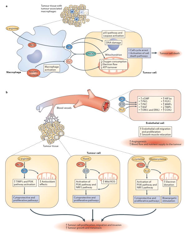
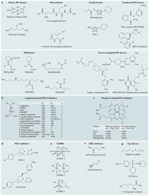
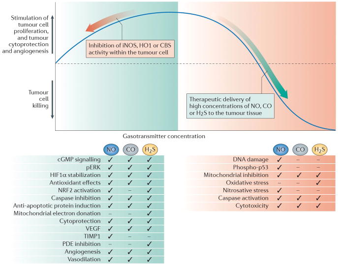
The three endogenous gaseous transmitters - nitric oxide (NO), carbon monoxide (CO) and hydrogen sulfide (H2S) - regulate a number of key biological functions. Emerging data have revealed several new mechanisms for each of these three gasotransmitters in tumour biology. It is now appreciated that they show bimodal pharmacological character in cancer, in that not only the inhibition of their biosynthesis but also elevation of their concentration beyond a certain threshold can exert anticancer effects. This Review discusses the role of each gasotransmitter in cancer and the effects of pharmacological agents - some of which are in early-stage clinical studies - that modulate the levels of each gasotransmitter. A clearer understanding of the pharmacological character of these three gases and the mechanisms underlying their biological effects is expected to guide further clinical translation.


Conflict of interest statement

Competing interests

The author declares competing interests: see Web version for details.

Figures

Figure 1
Figure 1. Effects of NO, CO and H2S on tumour survival and growth
[a | NO-mediated mechanisms of tumour cell killing by tumour-associated macrophages. Upregulation of inducible nitric oxide synthase (iNOS) in activated tumour-associated macrophages leads to the production of high local levels of NO. At the same time, macrophages also produce superoxide (O2) from nicotinamide adenine dinucleotide phosphate (NADPH) oxidase and other cellular sources. Together, NO and superoxide form peroxynitrite (ONOO), a reactive oxidant species. The resulting combination of nitrosative and oxidative stress can be cytostatic or cytotoxic to certain tumour cell types (the NO-associated component of cell killing is tumour-cell-type dependent). In susceptible tumour cells, the NO-mediated cell killing involves the inhibition of mitochondrial activity, DNA damage and activation of downstream pathways including such as the p53 and caspase activation pathways, culminating in tumour cell lysis. These mechanisms can be enhanced by various immunostimulatory therapies and/or by supplementation of L-arginine, the substrate of NOS. b | Pro-tumour effects of low levels of endogenously produced NO, carbon monoxide (CO) and hydrogen sulfide (H2S). Survival and proliferation of the tumour cell is stimulated by gasotransmitter production within the tumour. Within the tumour, induction of iNOS and consequently elevated levels of NO (top right), induction of haem oxygenase 1 (HO1) and elevated levels of CO (middle cancer cell on top of the right side of the graph) and/or induction of cystathionine-β-synthase (CBS) and elevated levels of H2S (bottom right) can exert pro-survival and pro-proliferative effects. Depending on the gasotransmitter, these signalling mechanisms can culminate in upregulation of fibroblast growth factor 2 (FGF2), activation of matrix metalloproteinases (MMPs), upregulation of tissue inhibitors of matrix metalloproteinases (TIMPs), activation of PI3K, and/or the stimulation of the inducible isoform of cyclooxygenase (COX2). In addition to tumour-autonomous effects, each gasotransmitter can diffuse out from the tumour cells and can stimulate intra- and peritumour angiogenesis through paracrine actions on endothelial cells, for instance by stimulating various pro-angiogenic pathways (including the cyclic GMP–protein kinase G (PKG) signalling pathway, activation of protein kinase C (PKC), RAF, extracellular signal-regulated kinase 1 (ERK1) and ERK2, and stabilization of hypoxia inducible factor 1α (HIF1α)). Although the signalling mechanisms are gasotransmitter- and condition-dependent, the ultimate result is the stimulation of peritumour angiogenesis and an increase in tumour blood flow.
Figure 2
Figure 2. Chemical structures of selected compounds that affect levels of gasotransmitters
a | Chemical structures of selected NO donor molecules. SNP and glyceryl trinitrate are considered “classic molecules”, which have been used by cardiologists for several decades. Nitrosothiols, syndonimines and NONOates have different half-lives/NO release profiles, but do not offer tumor-cell selectivity. The examples indicated in the figure are research compounds, rather than clinical development candidates. The “combined NO donors” (selected examples of which are shown here) offer the combined pharmacological action of the parent compound (e.g. a nonsteroidal anti-inflammatory) and the NO donating group; several members of this class are in various stages or preclinical or clinical development. The tumour-targeted NO donors utilise specific features of the tumour microenvironment to direct the release of NO within the tumour cells, in order to increase tumour cell specificity and to reduce potential systemic side effects of NO; these compounds are currently in preclinical testing. b | Chemical structures of selected L-arginine-based NOS inhibitors. In L-NAME, the acid functional group (-COO) becomes CO-O-CH3; **GW273629 is (3-[[2-[(1-iminoethyl)amino]ethyl]sulfonyl]-L-alanine); the central portion of the molecule contains a sulfonyl group within a 3-membered carbon chain; cindunistat is S-[2-(ethanimidoylamino)ethyl]-2-methyl- L-cysteine, where the central portion of the molecule contains a sulfur atom within a 3-membered carbon chain. c | Chemical structures of porphyrin-based haem oxygenase 1 (HO1) inhibitor compounds. Zinc, tin, manganese and chromium protoporphyrins have been described as competitive inhibitors for HO1 in the liver, spleen, kidney and other tissues. Most studies in cancer utilize ZnPP and SnPP. Sn-mesoporphyrin (Stanate; InfaCare Pharmaceutical Corporation) is noteworthy, as it has already been used in human studies. d | Chemical structures of selected non-porphyrin-based HO1 inhibitor compounds. OB24, an imidazole-dioxolane compound, is a member of a large group of compounds (that also contains imidazole ketones and imidazole alcohols) that are competitive inhibitors of HO1. OB24 has demonstrated efficacy in tumour-bearing mice models in vivo. Azalanstat is another potent HO1 inhibitor (IC50 values: 6 and 28 μM for rat HO1 and HO2, respectively). e | Chemical structures of selected CO-releasing molecules (CORMs). Each CORM molecule releases 1 molecule of CO; CORM1 and CORM2 releases CO rapidly (half-life: approx. 1 minute); CORM3 is a slower releaser of CO (half-life: approx. 1 hour). f | Chemical structures of two inhibitors of the hydrogen sulfide-producing enzyme cystathionine-β-synthase (CBS). G. Chemical structures of commonly used H2S donors.
Figure 3
Figure 3. The implications of the bell-shaped pharmacological profile of NO, CO and H2S for the therapy of cancer
Low concentrations of nitric oxide (NO), carbon monoxide (CO) and hydrogen sulfide (H2S) that are produced endogenously by inducible NO synthase (iNOS), haem oxygenase 1 (HO1) and cystathionine-β-synthase (CBS), respectively, can support tumour growth and tumour angiogenesis through the mediators and effects listed in the green box. Inhibition of these responses (depicted by the red arrow on the left side of the graph) can be of therapeutic benefit, either on its own, or to sensitize the tumour cell to standard anticancer therapies. High concentrations of the gasotransmitters can be cytostatic or cytotoxic; thus, therapeutic administration of each gasotransmitter (depicted by the green arrow on the right side of the graph), to sufficiently high concentrations in the tumour cell can be used to induce anticancer effects (listed in red box) and/or to potentiate anticancer chemo-or radiotherapy. Ticks indicate key pathways or mechanisms involved in the biological actions of low or high levels of each gasotransmitter. Please note that the figure incorporates some generalization; the pathways and mechanisms involved in the action of each gasotransmitter can be dependent on the cell-type and the experimental condition used in the various studies.

References

    1. Nathan C. Nitric oxide as a secretory product of mammalian cells. FASEB J. 1992;6:3051–3064. - PubMed
    1. Stuehr DJ. Mammalian nitric oxide synthases. Biochim Biophys Acta. 1999;1411:217–230. - PubMed
    1. Liaudet L, Soriano FG, Szabo C. Biology of nitric oxide signaling. Crit Care Med. 2000;28:N37–52. - PubMed
    1. Ignarro LJ, editor. Nitric Oxide, Biology and Pathobiology. 2. Academic Press; 2009.
    1. Southan GJ, Szabo C. Selective pharmacological inhibition of distinct nitric oxide synthase isoforms. Biochem Pharmacol. 1996;51:383–394. - PubMed

Publication types

MeSH terms