Review
. 2016 Mar;12(3):144-53.
doi: 10.1038/nrendo.2015.216. Epub 2015 Dec 18.

Insulin resistance and hyperinsulinaemia in diabetic cardiomyopathy

Affiliations
Review

Insulin resistance and hyperinsulinaemia in diabetic cardiomyopathy

Guanghong Jia et al. Nat Rev Endocrinol. 2016 Mar.

Abstract

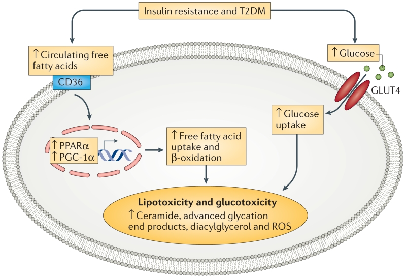
Insulin resistance, type 2 diabetes mellitus and associated hyperinsulinaemia can promote the development of a specific form of cardiomyopathy that is independent of coronary artery disease and hypertension. Termed diabetic cardiomyopathy, this form of cardiomyopathy is a major cause of morbidity and mortality in developed nations, and the prevalence of this condition is rising in parallel with increases in the incidence of obesity and type 2 diabetes mellitus. Of note, female patients seem to be particularly susceptible to the development of this complication of metabolic disease. The diabetic cardiomyopathy observed in insulin- resistant or hyperinsulinaemic states is characterized by impaired myocardial insulin signalling, mitochondrial dysfunction, endoplasmic reticulum stress, impaired calcium homeostasis, abnormal coronary microcirculation, activation of the sympathetic nervous system, activation of the renin-angiotensin-aldosterone system and maladaptive immune responses. These pathophysiological changes result in oxidative stress, fibrosis, hypertrophy, cardiac diastolic dysfunction and eventually systolic heart failure. This Review highlights a surge in diabetic cardiomyopathy research, summarizes current understanding of the molecular mechanisms underpinning this condition and explores potential preventive and therapeutic strategies.


Figures

Figure 1
Figure 1. Molecular mechanisms implicated in the development of diabetic cardiomyopathy
Overfeeding, mobilization of free fatty acids and activation of the renin–angiotensin–aldosterone system can all cause mitochondrial dysfunction, endoplasmic reticulum stress and oxidative stress, which results in impairment of insulin signalling, abnormal Ca2+ handling, increases in intracellular Ca2+ and Ca2+ sensitization increase and cardiomyocyte death. Patients eventually develop cardiomyocyte stiffness and diabetic cardiomyopathy. [Ca2+]i, Ca2+ influx; IRS-1, insulin receptor substrate 1; mTOR, mechanistic target of rapamycin (mTOR); P, phosphorylation; PI3K, phosphatidylinositol 3-kinase; Redox, oxidation–reduction state; S6K1, S6 kinase 1; Ser, serine; Thr, threonine; Tyr, tyrosine.
Figure 2
Figure 2. The development and progression of diabetic cardiomyopathy
Insulin resistance and hyperinsulinaemia increase systemic metabolic disorders, activate the SNS, activate RAAS, prompt oxidative stress, mitochondrial dysfunction and endoplasmic reticulum stress and impair calcium homeostasis. These effects result in cardiac fibrosis, hypertrophy, cardiomyocyte death, dysfunction of the coronary microcirculation and eventually heart failure. Furthermore, these pathophysiological changes in cardiomyocytes underlie the risk factors for insulin resistance and hyperinsulinaemia, which can result in a potentially vicious cycle. [Ca2+]i, Ca2+ influx; RAAS, renin–angiotensin–aldosterone system; SNS, sympathetic nervous system.
Figure 3
Figure 3. Insulin resistance and T2DM leads to system metabolic disorders in the cardiomyocyte
Under T2DM conditions, the expression of cardiac PPARα and PGC-1α is increased, which enhances the transcription of proteins that control free fatty acid uptake and oxidation. As glucose is a more efficient substrate than free fatty acids, a cardiac metabolic switch from glucose metabolism to free fatty acid oxidation decreases cardiac efficiency. This switch results in further metabolic disorders to cardiac dysfunction. CD36, cluster of differentiation 36; GLUT4, glucose transporter 4; T2DM, type 2 diabetes mellitus; PPARα; peroxisome proliferator activator receptor-α; PGC-1α, peroxisome proliferator-activated receptor gamma co-activator 1α; ROS, reactive oxygen species.

References

    1. Isfort M, Stevens SC, Schaffer S, Jong CJ, Wold LE. Metabolic dysfunction in diabetic cardiomyopathy. Heart Fail. Rev. 2014;19:35–48. - PMC - PubMed
    1. Adeghate E, Singh J. Structural changes in the myocardium during diabetes-induced cardiomyopathy. Heart Fail. Rev. 2014;19:15–23. - PubMed
    1. Dhalla NS, Takeda N, Rodriguez-Leyva D, Elimban V. Mechanisms of subcellular remodeling in heart failure due to diabetes. Heart Fail. Rev. 2014;19:87–99. - PubMed
    1. Velez M, Kohli S, Sabbah HN. Animal models of insulin resistance and heart failure. Heart Fail. Rev. 2014;19:1–13. - PMC - PubMed
    1. Jia G, Aroor AR, Martinez-Lemus LA, Sowers JR. Overnutrition, mTOR signaling, and cardiovascular diseases. Am. J. Physiol. Regul. Integr. Comp. Physiol. 2014;307:R1198–R1206. - PMC - PubMed

Publication types

MeSH terms