. 2016 Mar;37(3):1091-102.
doi: 10.1002/hbm.23088. Epub 2015 Dec 17.

Phasic and sustained brain responses in the amygdala and the bed nucleus of the stria terminalis during threat anticipation

Affiliations

Phasic and sustained brain responses in the amygdala and the bed nucleus of the stria terminalis during threat anticipation

Martin J Herrmann et al. Hum Brain Mapp. 2016 Mar.

Abstract

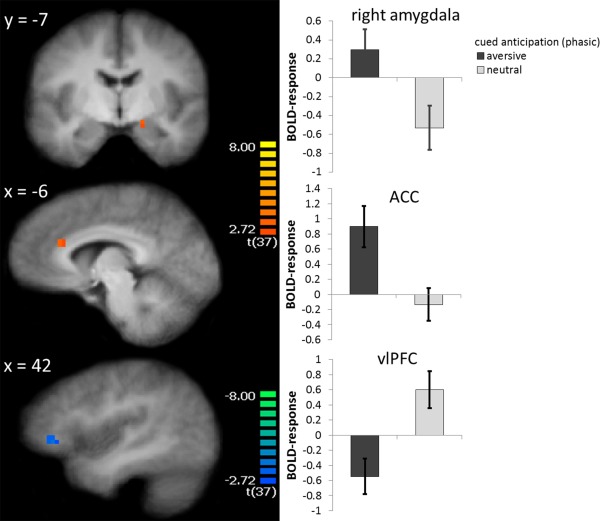
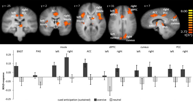
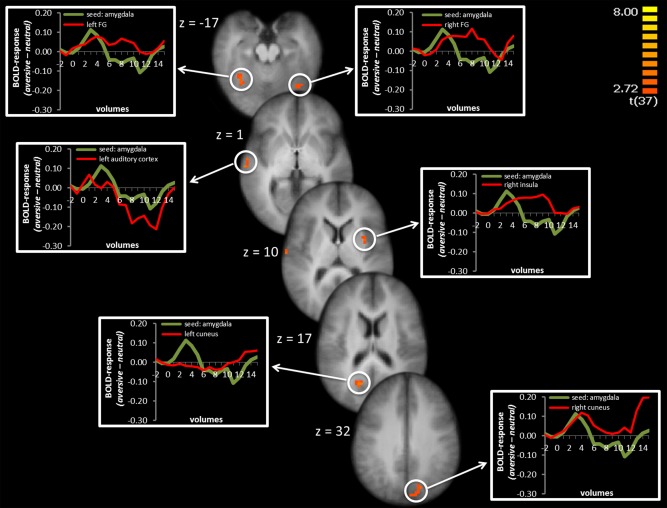
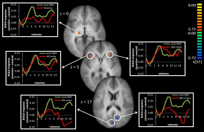
Several lines of evidence suggest that the amygdala and the bed nucleus of the stria terminalis (BNST) are differentially involved in phasic and sustained fear. Even though, results from neuroimaging studies support this distinction, a specific effect of a temporal dissociation with phasic responses to onset versus sustained responses during prolonged states of threat anticipation has not been shown yet. To explore this issue, we investigated brain activation during anticipation of threat in 38 healthy participants by means of functional magnetic resonance imaging. Participants were presented different visual cues indicated the temporally unpredictable occurrence of a subsequent aversive or neutral stimulus. During the onset of aversive versus neutral anticipatory cues, results showed a differential phasic activation of amygdala, anterior cingulate cortex (ACC), and ventrolateral prefrontal cortex (PFC). In contrast, activation in the BNST and other brain regions, including insula, dorsolateral PFC, ACC, cuneus, posterior cingulate cortex, and periaqueductal grey was characterized by a sustained response during the threat versus neutral anticipation period. Analyses of functional connectivity showed phasic amygdala response as positively associated with activation, mainly in sensory cortex areas whereas sustained BNST activation was negatively associated with activation in visual cortex and positively correlated with activation in the insula and thalamus. These findings suggest that the amygdala is responsive to the onset of cues signaling the unpredictable occurrence of a potential threat while the BNST in concert with other areas is involved in sustained anxiety. Furthermore, the amygdala and BNST are characterized by distinctive connectivity patterns during threat anticipation.

Keywords: phasic and sustained fear; fMRI; amygdala; BNST; insula.


Figures

Figure 1
Figure 1
Phasic fear: During the onset of anticipatory cues, participants showed increased activation to aversive vs. neutral cue onset in the right amygdala, anterior cingulate cortex (ACC), and decreased activation to aversive vs. neutral cue onset in the right ventrolateral prefrontal cortex (vlPFC). Statistical parametric maps are overlaid on an averaged T1 scan. The graphs on the right side display parameter estimates per condition (mean ± standard error for the maximally activated voxel). [Color figure can be viewed in the online issue, which is available at http://wileyonlinelibrary.com.]
Figure 2
Figure 2
Sustained fear: During the entire anticipation interval, participants showed greater activation in the right bed nucleus of stria terminalis (BNST), periaquaductal gray (PAG), left and right insula, anterior cingulate cortex (ACC), left and right dlPFC, cuneus, and posterior cingulate cortex (PCC) during the anticipation of aversive vs. neutral sounds. Statistical parametric maps are overlaid on an averaged T1 scan. The graph below displays parameter estimates per condition (mean ± standard error for the maximally activated voxel). [Color figure can be viewed in the online issue, which is available at http://wileyonlinelibrary.com.]
Figure 3
Figure 3
Psychophysiological interaction of right amygdala seed: Phasic activation in the right amygdala was positively coupled with activation in left and right fusiform gyrus (FG), left auditory cortex, right insula, and left and right cuneus. Significant voxels are overlaid on an averaged T1 scan. Time courses of activation are shown in the graphs at the left and right side starting two volumes before cue presentation and the first 15 volumes of cue presentation. The green line displays the difference between aversive‐neutral sound anticipation in the seed region and the red line display the difference between aversive‐neutral sound anticipation in the co‐activated brain region. [Color figure can be viewed in the online issue, which is available at http://wileyonlinelibrary.com.]
Figure 4
Figure 4
Psychophysiological interaction of right BNST seed: Sustained activation in the right BNST was positively coupled with activation in the left thalamus and left and right insula and negatively associated with activation in the left and right cuneus. Significant voxels are overlaid on an averaged T1 scan. Time courses of activation are shown in the graphs at the left and right side starting two volumes before cue onset. The green line displays the difference between aversive‐neutral sound anticipation in the seed region and the red line displays the difference between aversive‐neutral sound anticipation in the co‐activated brain region. [Color figure can be viewed in the online issue, which is available at http://wileyonlinelibrary.com.]

References

    1. Adhikari A (2014): Distributed circuits underlying anxiety. Front Behav Neurosci 8:112 - PMC - PubMed
    1. Alexander N, Osinsky R, Schmitz A, Mueller E, Kuepper Y, Hennig J (2010): The BDNF Val66Met polymorphism affects HPA‐axis reactivity to acute stress. Psychoneuroendocrinology 35:949–953. - PubMed
    1. Alvarez RP, Chen G, Bodurka J, Kaplan R, Grillon C (2011): Phasic and sustained fear in humans elicits distinct patterns of brain activity. NeuroImage 55:389–400. - PMC - PubMed
    1. Amaral DG, Behniea H, Kelly JL (2003): Topographic organization of projections from the amygdala to the visual cortex in the macaque monkey. Neuroscience 118:1099–1120. - PubMed
    1. Avery SN, Clauss JA, Winder DG, Woodward N, Heckers S, Blackford JU (2014): BNST neurocircuitry in humans. NeuroImage 91:311–323. - PMC - PubMed

Publication types