

LipidII: Just Another Brick in the Wall?
- PMID: 26679002
- PMCID: PMC4683061
- DOI: 10.1371/journal.ppat.1005213
LipidII: Just Another Brick in the Wall?
Abstract
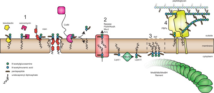
Nearly all bacteria contain a peptidoglycan cell wall. The peptidoglycan precursor molecule is LipidII, containing the basic peptidoglycan building block attached to a lipid. Although the suitability of LipidII as an antibacterial target has long been recognized, progress on elucidating the role(s) of LipidII in bacterial cell biology has been slow. The focus of this review is on exciting new developments, both with respect to antibacterials targeting LipidII as well as the emerging role of LipidII in organizing the membrane and cell wall synthesis. It appears that on both sides of the membrane, LipidII plays crucial roles in organizing cytoskeletal proteins and peptidoglycan synthesis machineries. Finally, the recent discovery of no less than three different categories of LipidII flippases will be discussed.
Conflict of interest statement
The authors have declared that no competing interests exist.
Figures
Similar articles
-
The localization of key Bacillus subtilis penicillin binding proteins during cell growth is determined by substrate availability.Environ Microbiol. 2013 Dec;15(12):3272-81. doi: 10.1111/1462-2920.12206. Epub 2013 Jul 30. Environ Microbiol. 2013. PMID: 23895585
-
Recent Advances in Peptidoglycan Synthesis and Regulation in Bacteria.Biomolecules. 2023 Apr 22;13(5):720. doi: 10.3390/biom13050720. Biomolecules. 2023. PMID: 37238589 Free PMC article. Review.
-
Cutting and stitching: the cross-linking of peptidoglycan in the assembly of the bacterial cell wall.ACS Chem Biol. 2007 Sep 21;2(9):602-5. doi: 10.1021/cb700182u. ACS Chem Biol. 2007. PMID: 17894443 Review.
-
Regulation of peptidoglycan synthesis and remodelling.Nat Rev Microbiol. 2020 Aug;18(8):446-460. doi: 10.1038/s41579-020-0366-3. Epub 2020 May 18. Nat Rev Microbiol. 2020. PMID: 32424210 Review.
-
Targeting the Achilles' Heel of Bacteria: Different Mechanisms To Break Down the Peptidoglycan Cell Wall during Bacterial Warfare.J Bacteriol. 2021 Mar 8;203(7):e00478-20. doi: 10.1128/JB.00478-20. Print 2021 Mar 8. J Bacteriol. 2021. PMID: 33139480 Free PMC article. Review.
Cited by
-
The evolution of spherical cell shape; progress and perspective.Biochem Soc Trans. 2019 Dec 20;47(6):1621-1634. doi: 10.1042/BST20180634. Biochem Soc Trans. 2019. PMID: 31829405 Free PMC article. Review.
-
XAC4296 Is a Multifunctional and Exclusive Xanthomonadaceae Gene Containing a Fusion of Lytic Transglycosylase and Epimerase Domains.Microorganisms. 2022 May 11;10(5):1008. doi: 10.3390/microorganisms10051008. Microorganisms. 2022. PMID: 35630451 Free PMC article.
-
Direct observation of the influence of cardiolipin and antibiotics on lipid II binding to MurJ.Nat Chem. 2018 Mar;10(3):363-371. doi: 10.1038/nchem.2919. Epub 2018 Jan 8. Nat Chem. 2018. PMID: 29461535 Free PMC article.
-
Cell Cycle Machinery in Bacillus subtilis.Subcell Biochem. 2017;84:67-101. doi: 10.1007/978-3-319-53047-5_3. Subcell Biochem. 2017. PMID: 28500523 Free PMC article. Review.
-
Single-cell growth inference of Corynebacterium glutamicum reveals asymptotically linear growth.Elife. 2021 Oct 4;10:e70106. doi: 10.7554/eLife.70106. Elife. 2021. PMID: 34605403 Free PMC article.
References
Publication types
MeSH terms
Substances
LinkOut - more resources
Full Text Sources
Other Literature Sources
