. 2016 Feb 26;291(9):4356-73.
doi: 10.1074/jbc.M115.700385. Epub 2015 Dec 17.

Selective Inhibition of the Mitochondrial Permeability Transition Pore Protects against Neurodegeneration in Experimental Multiple Sclerosis

Affiliations

Selective Inhibition of the Mitochondrial Permeability Transition Pore Protects against Neurodegeneration in Experimental Multiple Sclerosis

Justin Warne et al. J Biol Chem. .

Abstract

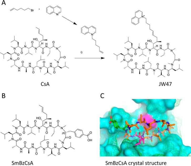
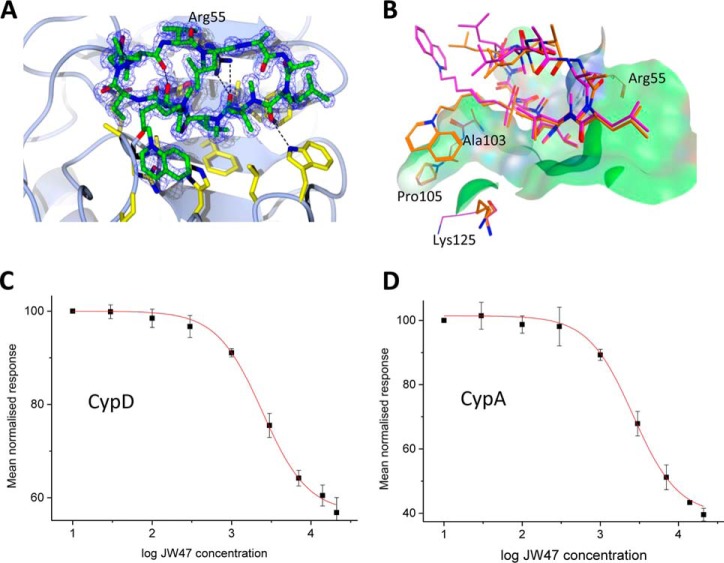
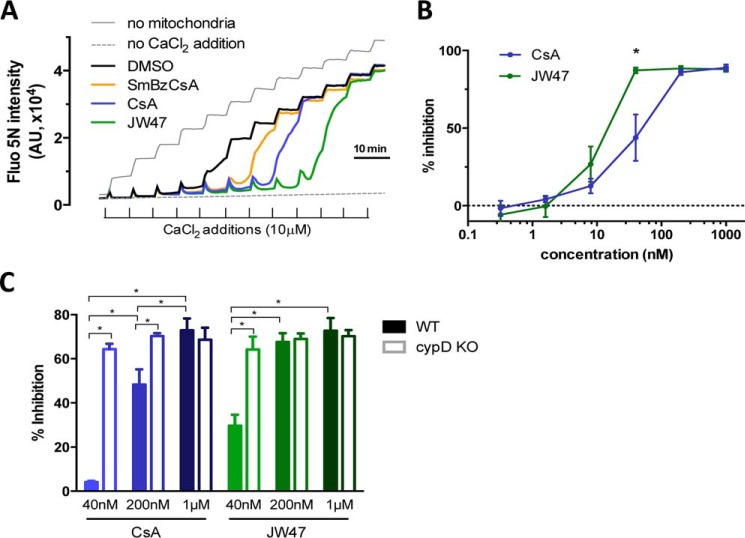
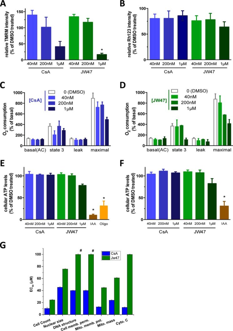
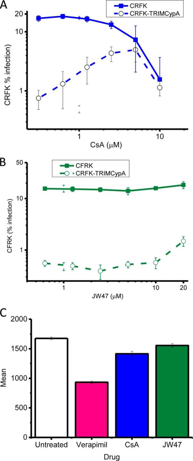
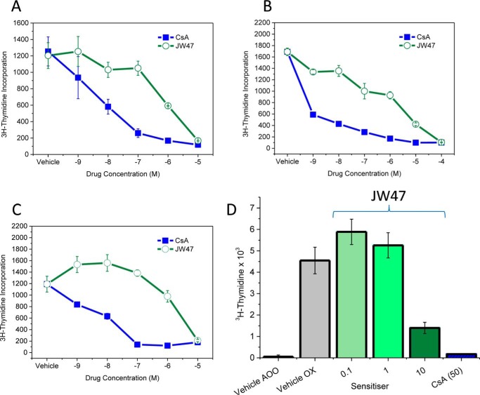
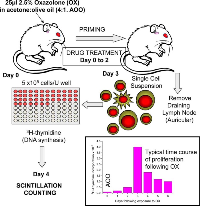
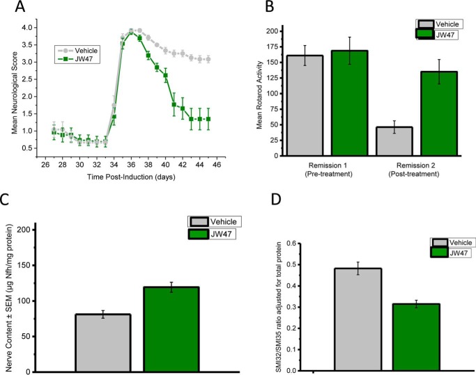
The mitochondrial permeability transition pore is a recognized drug target for neurodegenerative conditions such as multiple sclerosis and for ischemia-reperfusion injury in the brain and heart. The peptidylprolyl isomerase, cyclophilin D (CypD, PPIF), is a positive regulator of the pore, and genetic down-regulation or knock-out improves outcomes in disease models. Current inhibitors of peptidylprolyl isomerases show no selectivity between the tightly conserved cyclophilin paralogs and exhibit significant off-target effects, immunosuppression, and toxicity. We therefore designed and synthesized a new mitochondrially targeted CypD inhibitor, JW47, using a quinolinium cation tethered to cyclosporine. X-ray analysis was used to validate the design concept, and biological evaluation revealed selective cellular inhibition of CypD and the permeability transition pore with reduced cellular toxicity compared with cyclosporine. In an experimental autoimmune encephalomyelitis disease model of neurodegeneration in multiple sclerosis, JW47 demonstrated significant protection of axons and improved motor assessments with minimal immunosuppression. These findings suggest that selective CypD inhibition may represent a viable therapeutic strategy for MS and identify quinolinium as a mitochondrial targeting group for in vivo use.

Keywords: EAE; X-ray crystallography; cyclophilin D; cyclosporin; mitochondrial permeability transition (MPT); mitochondrial targeting; multiple sclerosis; neurodegeneration; neurodegenerative disease.


Figures

FIGURE 1.
FIGURE 1.
A, synthesis of [Bmt1]- and [Sar3]-substituted analogues by olefin cross-metathesis and alkylation. Reagents were DCM, Hoveyda-Grubbs catalyst, second generation, reflux, 30 h (i) and ethyl acetate reflux (ii). B, structure of SmBzCsA. C, the crystal structure of SmBzCsA (Protein Data Bank code 4IPZ) is shown bound to CypA (solid magenta surface). The aligned structure of CypD (Protein Data Bank code 5A0E) is shown as a wire mesh. Amino acids 9 to 2 are involved in cyclophilin binding. The [Bmt1] residue (pink) side chain and the [Sar3] substituent do not affect cyclophilin binding.
FIGURE 2.
FIGURE 2.
A, crystal structure of JW47 in complex with CypD and its effect on Ca2+-induced PT. Electron density of JW47 in the CypD catalytic site is shown as a wire mesh with JW47 modeled into the density as a ball and stick model (green). B, JW47 adopts two poses in the crystal structure shown in orange and magenta. The orange pose illustrates a possible stabilizing interaction with Ala-103, Pro-105, and Lys-125. The surface in green is generated from the chain that co-crystallized with this pose. Note that Lys-125 also adopts two conformations. C, fluorescence polarization assay for cyclophilin binding obtained using a fluorescein-labeled CsA probe. Typical data for CypD are shown. The concentration of the probe is 45 nm, and the enzyme concentration is 40 nm. Millipolarization values are fitted to a dose-response curve using Origin. D, fluorescence polarization data for CypA. Error bars, S.D.
FIGURE 3.
FIGURE 3.
A, comparison of the effect of JW47, CsA, and SmBzCsA on mitochondrial CRC. Shown are representative traces of the CRC assay in isolated rat liver mitochondria. Fluo-5N fluorescence was measured in the extramitochondrial solution following repeated additions of Ca2+ (10 μm; for details see “Experimental Procedures”). Increase in fluorescence indicates loss of Ca2+ retention due to PT pore opening. Inhibition of PT pore with different compounds (added at 200 nm) increases CRC, represented by the delay in increase of fluorescence. B, dose-response curves of JW47 and CsA on PT inhibition (expressed as percentage increase in CRC compared with DMSO treatment). C, quantification of the dose-dependent effects of CsA and JW47 on CRC (PT) in liver mitochondria isolated from WT and CypD KO animals. Percentage inhibition denotes increase in CRC compared with DMSO treatment, normalized to WT. *, p < 0.05 (t test). Error bars, S.E.
FIGURE 4.
FIGURE 4.
Mitochondrial and cellular toxicological assessment of JW47 and CsA. Mitochondrial parameters were measured in rat cortical neurons (A and E) and isolated rat liver mitochondria (B–D and F). A and B, mitochondrial membrane potential was measured in tetramethylrhodamine methyl ester (TMRM)-loaded neurons using ImageXpress MicroXL (A) and in rhodamine-123 loaded isolated mitochondria using a fluorescent plate reader (B). Values are normalized to DMSO (100%)- and FCCP (2.5 μm, 0%)-treated samples. *, p < 0.05 (one-way ANOVA). C and D, O2 consumption was measured in mitochondria isolated from rat liver in the presence of glutamate and malate using an Oroboros high resolution oxygraph as described under “Experimental Procedures.” The effect of compounds on basal, leak (oligomycin, 2.5 μm), and maximal uncoupled respiration (FCCP, titrated to give maximum effect) is shown, as compared with basal, DMSO controls. *, p < 0.05 (paired t tests). E and F, ATP levels in cortical neurons (E) and ATP production of isolated mitochondria in the presence of substrates and ADP (F) was measured using a luciferase assay as described under “Experimental Procedures.” Iodoacetic acid (IAA; 1 mm) and oligomycin (oligo; 2.5 μm) were used to show the contribution of glycolysis and mitochondrial ATP synthesis, respectively. *, p < 0.05 (t test). G, in vitro toxicological assessment of CsA and JW47. HepG2 cells were plated, and after 24 h, the cells were treated with the compounds at a range of concentrations. At the end of the incubation period, the cells were loaded with the relevant dye/antibody for each cell health marker and scanned (see “Experimental Procedures”). Data are shown as EC50 values (μm) ± R2. #, no response observed at 100 μm. Error bars, S.E.
FIGURE 5.
FIGURE 5.
Assessment of in-cell CypA binding. CRFK cells transduced with either empty vector (filled squares) or TRIM-CypA (open circles) were infected with VSV-pseudotyped GFP-expressing HIV-1 vector in the presence of DMSO or increasing concentration of drug. A, CsA; B, JW47. Viral infection was measured by flow cytometry at 48 h postinfection. Data are the average of three independent experiments. C, P-glycoprotein drug transporter activity. Vehicle, verapamil (100 μm), and CsA analogues were tested as substrates for P-glycoprotein (Pgp-Glo assay). Results are the mean ± S.E. (error bars) bioluminescence measurements from the luciferin reporter.
FIGURE 6.
FIGURE 6.
The oxazolone contact hypersensitivity test. A low severity in vivo measure of T cell proliferation is used as a rapid screen for immunosuppressive doses of agents. Oxazolone administered to the ear induces an increase in cell number and proliferation in the draining auricular lymph node. Error bars, S.E.
FIGURE 7.
FIGURE 7.
JW47 exhibits less immunosuppressive activity than CsA. Mitogenesis in vitro of normal mouse splenocyte cells is shown. Splenocytes were incubated with 5 μg/ml concanavalin A (A), mitogenic CD3/CD28 monoclonal antibodies (B), or splenocytes from MOG residue 35–55 peptide-immunized mice (C) in the presence of 5 μg/ml MOG peptide with vehicle or compounds for 2 (A and B) or 4 (C) days prior to the addition of 1 μCi of [3H]thymidine and were harvested 16–20 h later, and tritiated thymidine incorporation was assessed by β-scintillation counting. The results represent the mean ± S.E. of triplicate samples. D, low doses of JW47 in vivo exhibited no immunosuppressive activity. 25 μl of 2.5% oxazolone (OX) or acetone/olive oil (4:1) vehicle (AOO) was applied to the ear skin of ABH mice on day 0. On day 3, the draining auricular lymph nodes of 3–4 mice/group were removed, and 5 × 10⁵ cells were cultured overnight in the presence of 1 μCi of [3H]thymidine. Animals were treated with 0.1 ml of vehicle, 0.1–10 mg/kg JW47, or 50 mg/kg CsA. The results represent the mean ± S.E. of at least quadruplicate samples.
FIGURE 8.
FIGURE 8.
JW47 limits the accumulation of neurodegeneration and disability during relapsing autoimmune encephalomyelitis. ABH mice were injected with SCH in Freund's complete adjuvant on days 0 and 7 to induce paralytic EAE. A relapse was induced on day 28 during the first remission, using a further injection of SCH in Freund's incomplete adjuvant. Animals were treated daily from day 33 onward with either 1 mg/kg intraperitoneal JW47 (n = 14) in ethanol/cremophor/PBS (1:1:18) or vehicle (n = 12). The results represent the mean daily neurological score ± S.E. (error bars) (A) and the mean rotarod activity representing the mean ± S.E. time before falling/failing to stay on an accelerating rotarod before (on day 27) or after (on day 45) treatment with either vehicle or JW47 (B). *** p < 0.001 compared with vehicle treatment. C, axonal content in the spinal cord following treatment of relapsing EAE with JW47 1 mg/kg measured as neurofilament level adjusted for total protein content. EAE was induced with spinal cord homogenate in complete Freund's adjuvant on days 0 and 7, and a relapse was induced by reimmunization with spinal cord homogenate in complete Freund's adjuvant at day 28. Animals were randomized according to RotaRod performance score at day 27 to receive either vehicle (cremophor (Sigma)/alcohol/phosphate-buffered saline, 1:1:18) or 1 mg/kg intraperitoneal JW47 from day 33 postinfection just prior to the development of relapse at day 35 until day 47. Animals were killed, and the spinal cords were removed using hydrostatic pressure, and axonal content was measured using a quantitative neurofilament-specific ELISA. n = 11 untreated, n = 13 JW47-treated. Shown is the ratio of dephosphorylated (SMI32-reactive) neurofilament to hyperphosphorylated (SMI35-reactive) neurofilament as measured by ELISA in spinal cord homogenates from postrelapse untreated animals (n = 11) or JW47 (1 mg/kg)-treated animals (n = 13); ***, p < 0.001 adjusted for total protein level.

References

    1. Giorgio V., von Stockum S., Antoniel M., Fabbro A., Fogolari F., Forte M., Glick G. D., Petronilli V., Zoratti M., Szabó I., Lippe G., and Bernardi P. (2013) Dimers of mitochondrial ATP synthase form the permeability transition pore. Proc. Natl. Acad. Sci. U.S.A. 110, 5887–5892 - PMC - PubMed
    1. Alavian K. N., Beutner G., Lazrove E., Sacchetti S., Park H. A., Licznerski P., Li H., Nabili P., Hockensmith K., Graham M., Porter G. A. Jr., and Jonas E. A. (2014) An uncoupling channel within the c-subunit ring of the F1FO ATP synthase is the mitochondrial permeability transition pore. Proc. Natl. Acad. Sci. U.S.A. 111, 10580–10585 - PMC - PubMed
    1. Bonora M., Bononi A., De Marchi E., Giorgi C., Lebiedzinska M., Marchi S., Patergnani S., Rimessi A., Suski J. M., Wojtala A., Wieckowski M. R., Kroemer G., Galluzzi L., and Pinton P. (2013) Role of the c subunit of the FO ATP synthase in mitochondrial permeability transition. Cell Cycle 12, 674–683 - PMC - PubMed
    1. Halestrap A. P., and Richardson A. P. (2015) The mitochondrial permeability transition: a current perspective on its identity and role in ischaemia/reperfusion injury. J. Mol. Cell. Cardiol. 78, 129–141 - PubMed
    1. Giorgio V., Bisetto E., Soriano M. E., Dabbeni-Sala F., Basso E., Petronilli V., Forte M. A., Bernardi P., and Lippe G. (2009) Cyclophilin D modulates mitochondrial F0F1-ATP synthase by interacting with the lateral stalk of the complex. J. Biol. Chem. 284, 33982–33988 - PMC - PubMed

Publication types

MeSH terms