. 2016 Feb 12;291(7):3581-94.
doi: 10.1074/jbc.M115.689091. Epub 2015 Dec 17.

Structural Correlation of the Neck Coil with the Coiled-coil (CC1)-Forkhead-associated (FHA) Tandem for Active Kinesin-3 KIF13A

Affiliations

Structural Correlation of the Neck Coil with the Coiled-coil (CC1)-Forkhead-associated (FHA) Tandem for Active Kinesin-3 KIF13A

Jinqi Ren et al. J Biol Chem. .

Abstract

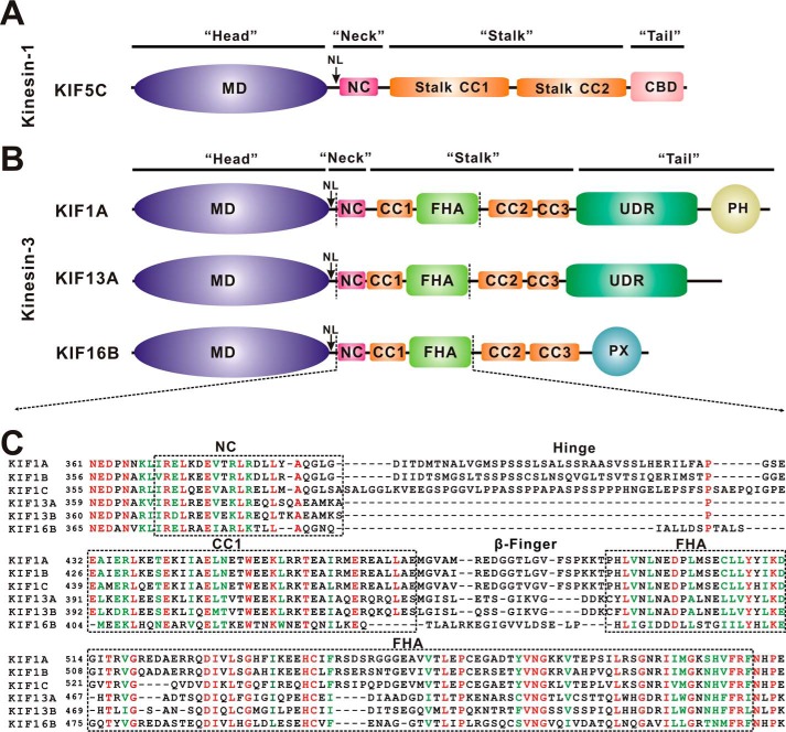
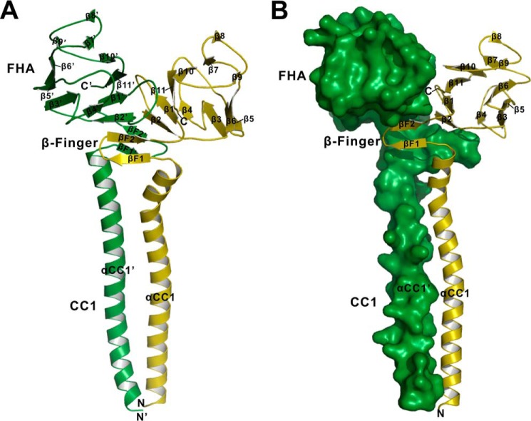
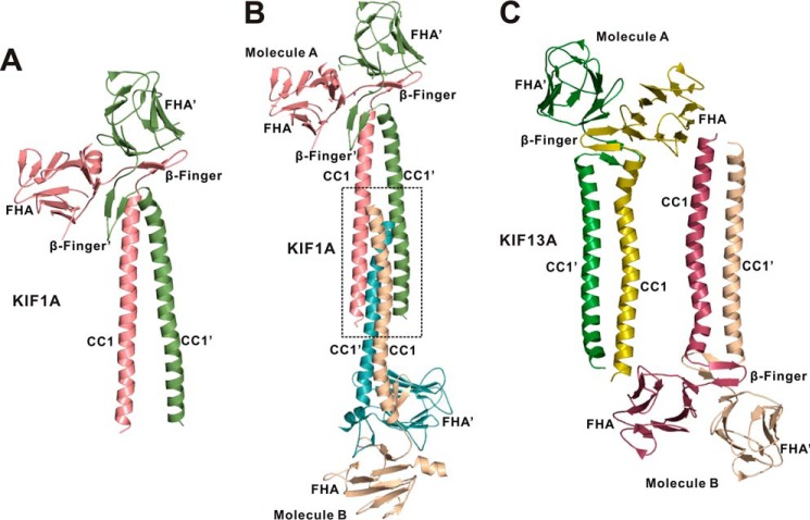
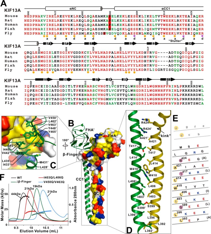
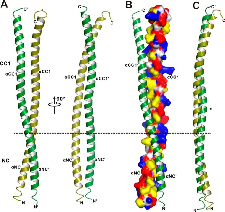
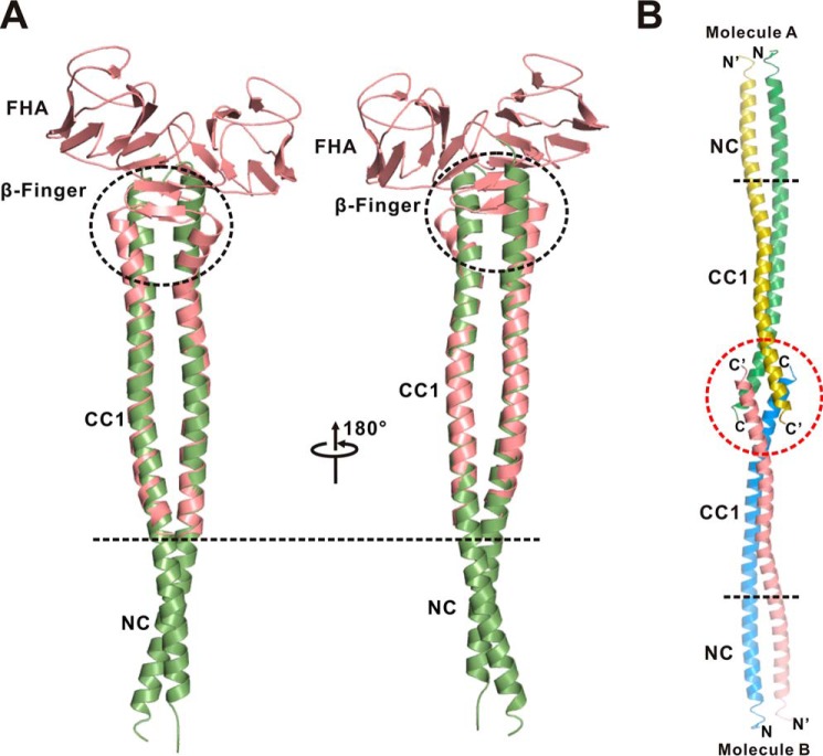
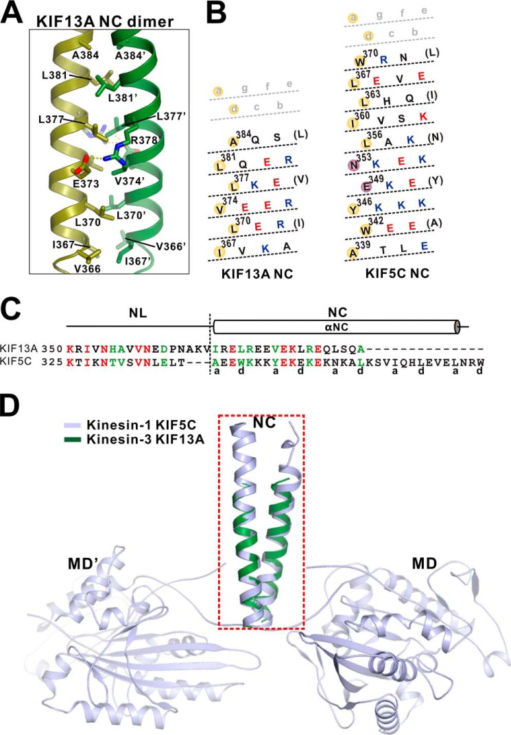
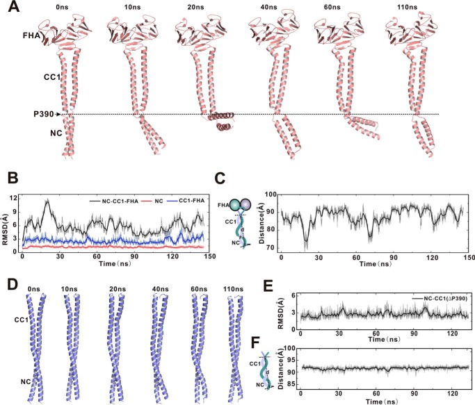
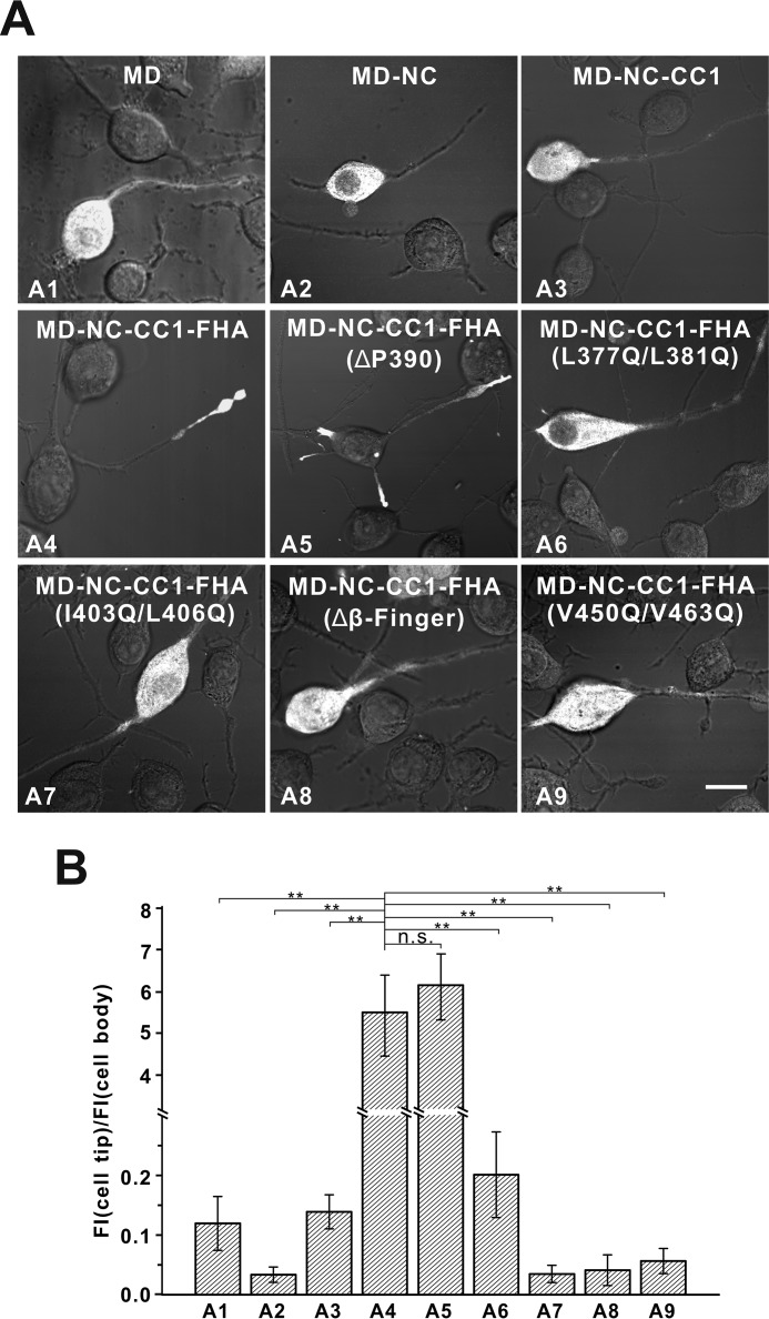
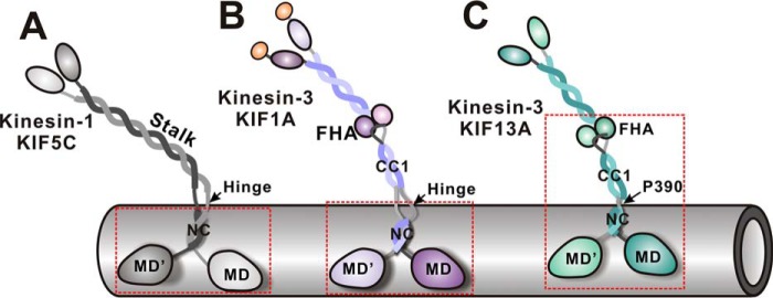
Processive kinesin motors often contain a coiled-coil neck that controls the directionality and processivity. However, the neck coil (NC) of kinesin-3 is too short to form a stable coiled-coil dimer. Here, we found that the coiled-coil (CC1)-forkhead-associated (FHA) tandem (that is connected to NC by Pro-390) of kinesin-3 KIF13A assembles as an extended dimer. With the removal of Pro-390, the NC-CC1 tandem of KIF13A unexpectedly forms a continuous coiled-coil dimer that can be well aligned into the CC1-FHA dimer. The reverse introduction of Pro-390 breaks the NC-CC1 coiled-coil dimer but provides the intrinsic flexibility to couple NC with the CC1-FHA tandem. Mutations of either NC, CC1, or the FHA domain all significantly impaired the motor activity. Thus, the three elements within the NC-CC1-FHA tandem of KIF13A are structurally interrelated to form a stable dimer for activating the motor. This work also provides the first direct structural evidence to support the formation of a coiled-coil neck by the short characteristic neck domain of kinesin-3.

Keywords: KIF13A; intracellular trafficking; kinesin; molecular motor; neck coil; structural biology; x-ray crystallography.


Figures

FIGURE 1.
FIGURE 1.
Domain organization of kinesin-1 and kinesin-3. A, domain organization of kinesin-1 KIF5C. KIF5C contains an N-terminal MD, a neck coil (NC), the central stalk CC1 and CC2, and a C-terminal cargo binding domain (CBD). B, domain organization of kinesin-3 KIF1A, KIF13A, and KIF16B. In addition to an N-terminal motor domain and a neck coil, KIF1A, KIF13A, and KIF16B contain non-continuous coiled-coils (CC1-CC3) and an FHA domain in the middle. Both KIF1A and KIF13A contain an undefined region (UDR), and KIF1A and KIF16B contain an extra PH domain and PX domain at the C terminus, respectively. C, sequence alignment of the NC-CC1-FHA tandem from different kinesin-3 motors. The identical residues are colored in red, and the highly conserved residues are colored in green. The regions of NC, CC1, and the FHA domain are marked with dashed boxes.
FIGURE 2.
FIGURE 2.
The overall structure of the CC1-FHA dimer of KIF13A. A, a ribbon diagram of the structure of the CC1-FHA dimer. The two subunits of the CC1-FHA dimer are colored in green and olive, respectively. The secondary structures of the FHA domain, the β-finger, and the CC1 helix are labeled, and both the N and C termini are also marked. B, a combined surface and ribbon representation of the dimer structure. One of the two subunits is in the surface representation (colored in green), and the other is in the ribbon representation (colored in olive).
FIGURE 3.
FIGURE 3.
Analysis of the crystal packing of the CC1-FHA dimers of KIF1A and KIF13A. A, a ribbon diagram of the structure of the CC1-FHA dimer of KIF1A (PDB code 4EGX). The two subunits of the dimer are colored in green and red, respectively. B, analysis of the crystal stacking of the CC1-FHA dimer of KIF1A. In the crystal stacking the two CC1 helices from one dimer (Molecule A) pack with the ones from the other dimer (Molecule B) to form a four-helix bundle, which induces the separation of the N-terminal halves of the two CC1 helices. C, analysis of the crystal stacking of the CC1-FHA dimer of KIF13A. In the crystal stacking, the two CC1 helices from one dimer (molecule A) do not interfere with the ones from the other dimer (molecule B).
FIGURE 4.
FIGURE 4.
The interaction interface between the CC1-FHA dimer. A, structure-based sequence alignment of the NC-CC1-FHA tandem of KIF13A from different species. The identical residues are colored in red, and the highly conserved residues are in green. The secondary structures and residue numbers are marked on the top. The hydrophobic residues responsible for the formation of the NC-CC1(ΔPro-390) and CC1-FHA dimers are highlighted by yellow circles at the bottom. The hydrophilic residues that are located in the interhelical packing between the CC1 dimer are marked by purple asterisks. The a and d sites of the NC and CC1 helices for interhelical packing are also marked. B, a combined surface and ribbon representation of the CC1-FHA dimer. In this surface drawing the hydrophobic, positively charged, negatively charged residues and remaining residues are colored in yellow, blue, red, and white, respectively. The interaction interface between the dimer can be divided into two parts (highlighted by boxes). C, a combined surface, ribbon and stick model illustrates the dimer interface mediated by the FHA domain and β-finger. The side chains of the residues involved in the dimer packing are shown as sticks. D, a combined ribbon and stick model illustrates the dimer interface mediated by CC1. The side chains of the residues involved in the dimer packing are shown as sticks. E, heptad repeat register of the residues for the CC1 dimer packing. The CC1 helix has been cut and opened flat to give a two-dimensional representation. The hydrophobic residues and hydrophilic residues in the packing core are highlighted by yellow and red circles, respectively. F, size exclusion chromatography-multiangle light scattering analysis of the CC1-FHA tandem and its mutants. The calculated molecular mass of each fragment is marked.
FIGURE 5.
FIGURE 5.
The structure of the NC-CC1(ΔPro-390) dimer. A, a ribbon diagram of the structure of the NC-CC1(ΔPro-390) dimer. The two subunits of the dimer are colored in green and olive, respectively. The division site between NC and CC1 is highlighted by a dashed line. The secondary structures of NC and CC1 are labeled, and the N and C termini are also marked. B, a combined surface and ribbon representation of the NC-CC1(ΔPro-390) dimer. In this surface drawing the color schemes follow that of Fig. 4B. C, superposition of the two subunits of the NC-CC1(ΔPro-390) dimer. Notably, one helix is slightly twisted in the middle (marked with an arrow).
FIGURE 6.
FIGURE 6.
The extended conformation of the NC-CC1-FHA(ΔPro-390) dimer of KIF13A. A, a structural model of the NC-CC1-FHA(ΔPro-390) dimer built by superimposing the structures of the NC-CC1(ΔPro-390) (green) and CC1-FHA (red) dimers based on CC1. The two CC1 dimers from the two structures can be well aligned with each other except for the extreme C-terminal ends (highlighted by a black circle). B, analysis of the crystal stacking of the NC-CC1(ΔPro-390) dimer of KIF13A. In the crystal stacking the two C-terminal ends of CC1 from one dimer (molecule A) pack with the ones from the other dimer (molecule B), which leads to the difference of the C-terminal end of CC1 between the two dimers.
FIGURE 7.
FIGURE 7.
The interhelical packing between the NC coiled-coil dimer. A, a combined ribbon and stick model illustrates the interaction interface between the NC coiled-coil dimer. The side chains of the residues involved in the interhelical packing are shown as sticks. B, heptad repeat register of the residues for the coiled-coil packing of the NC dimer of KIF13A and KIF5C. The representation mode and color scheme follow that of Fig. 4E. C, structure-based sequence alignment of the neck domain (NL and NC) of kinesin-3 KIF13A and kinesin-1 KIF5C. The identical residues are colored in red, and the highly conserved residues are colored in green. The a and d sites of the NC coiled-coil are marked at the bottom. D, superposition of the NC coiled-coil structure of KIF13A with that of KIF5C (PDB code 3KIN). The NC coiled-coil dimer of KIF13A (colored in green) can be well aligned with that of KIF5C (colored in light blue) but is much shorter.
FIGURE 8.
FIGURE 8.
Molecular dynamics simulations of the NC-CC1-FHA and NC-CC1(ΔPro-390) dimers. A, snapshots of a representative simulation of the NC-CC1-FHA dimer with simulation time indicated. The extended NC-CC1-FHA dimer is broken into two linked dimers (the NC dimer and the CC1-FHA dimer) by Pro-390 that is highlighted by a dashed line. B, time course of the root mean square deviation of the NC dimer (red), the CC1-FHA dimer (blue), and the NC-CC1-FHA dimer (black). Both the NC dimer and the CC1-FHA dimer undergo little conformational changes, but the overall conformation of the NC-CC1-FHA dimer is dynamic during simulations. RMSD, root mean square deviation. C, time course of the distance between the N-terminal end of NC and the C-terminal end of CC1 (as indicated in the left panel), indicating the significant fluctuations of the overall conformation of the NC-CC1-FHA dimer. D, snapshots of a representative simulation of the NC-CC1(ΔPro-390) dimer with simulation time indicated. E, time course of the root mean square deviation of the NC-CC1(ΔPro-390) dimer. F, time course of the distance between the N-terminal end of NC and the C-terminal end of CC1, indicating no significant conformational changes of the NC-CC1(ΔPro-390) dimer.
FIGURE 9.
FIGURE 9.
The NC-CC1-FHA tandem is essential for motor activation. A, cellular localizations of the MD, MD-NC, MD-NC-CC1, and MD-NC-CC1-FHA fragments and various MD-NC-CC1-FHA mutants. The MD, MD-NC, and MD-NC-CC1 fragments were all enriched in the cell body (A1–A3), whereas the MD-NC-CC1-FHA fragment was predominantly localized to the cell periphery (A4). The MD-NC-CC1-FHA mutants with the mutations to disrupt the dimer were localized in the cell body (A6–A9), but the NC-CC1-FHA(ΔPro-390) mutant was localized to the cell periphery (A5). Scale bar: 20 μm. B, quantification of the cellular distribution data shown in panel A. The ratio of the tip to cell body average fluorescence intensity (FI) was quantified for each construct for more than 15 cells (n > 15). Each bar represents the mean value ± S.D. **, p < 0.05. n.s., not significant.
FIGURE 10.
FIGURE 10.
Schematic models illustrating the active states of kinesin-1 and kinesin-3. In the active state of kinesin-1 KIF5C, the NC dimer alone is sufficient to activate the motor domain and the flexible linker between the neck and stalk is essential for processive movement (A). In contrast, the NC dimer of kinesin-3 KIF1A is unable to activate the motor domain and needs the subsequent segments (i.e. a part of the NC/CC1-hinge) to stabilize the dimer for motor activation (B). Due to the lack of the NC/CC1 hinge, the whole CC1-FHA tandem of KIF13A is required for correlating with the NC dimer to activate the motor domain (C). The minimum active fragments of KIF5C, KIF1A, and KIF13A are marked with dashed boxes. The intrinsic flexibility between the neck and stalk of kinesin-3 (provided by the NC/CC1-hinge of KIF1A and the Pro-390 linker of KIF13A) is also likely to be essential for active transport.

References

    1. Vale R. D. (2003) The molecular motor toolbox for intracellular transport. Cell 112, 467–480 - PubMed
    1. Soldati T., and Schliwa M. (2006) Powering membrane traffic in endocytosis and recycling. Nat. Rev. Mol. Cell Biol. 7, 897–908 - PubMed
    1. Hirokawa N., Niwa S., and Tanaka Y. (2010) Molecular motors in neurons: transport mechanisms and roles in brain function, development, and disease. Neuron 68, 610–638 - PubMed
    1. Hirokawa N., Noda Y., Tanaka Y., and Niwa S. (2009) Kinesin superfamily motor proteins and intracellular transport. Nat. Rev. Mol. Cell Biol. 10, 682–696 - PubMed
    1. Verhey K. J., Kaul N., and Soppina V. (2011) Kinesin assembly and movement in cells. Annu. Rev. Biophys. 40, 267–288 - PubMed

Publication types

MeSH terms

LinkOut - more resources