Cerebral Hypoperfusion in Hereditary Coproporphyria (HCP): A Single Photon Emission Computed Tomography (SPECT) Study
- PMID: 26680773
- PMCID: PMC5171194
- DOI: 10.2174/1871530316666151218151101
Cerebral Hypoperfusion in Hereditary Coproporphyria (HCP): A Single Photon Emission Computed Tomography (SPECT) Study
Abstract
Background: Hereditary Coproporphyria (HCP) is characterized by abdominal pain, neurologic symptoms and psychiatric disorders, even if it might remain asymptomatic. The pathophysiology of both neurologic and psychiatric symptoms is not fully understood. Therefore, aiming to evaluate a possible role of brain blood flow disorders, we have retrospectively investigated cerebral perfusion patterns in Single Photon Emission Computed Tomography (SPECT) studies in HCP patients.
Materials & methods: We retrospectively evaluated the medical records of patients diagnosed as being affected by HCP. A total of seven HCP patients had been submitted to brain perfusion SPECT study with 99mTc-Exametazime (hexamethylpropyleneamine oxime, HMPAO) or with its functionally equivalent 99mTc-Bicisate (ECD or Neurolite) according with common procedures. In 3 patients the scintigraphic study had been repeated for a second time after the first evaluation at 3, 10 and 20 months, respectively. All the studied subjects had been also submitted to an electromyographic and a Magnetic Resonance Imaging (MRI) study of the brain.
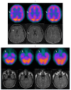
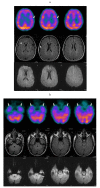
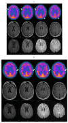
Results: Mild to moderate perfusion defects were detected in temporal lobes (all 7 patients), frontal lobes (6 patients) and parietal lobes (4 patients). Occipital lobe, basal ganglia and cerebellar involvement were never observed. In the three subjects in which SPECT study was repeated, some recovery of hypo-perfused areas and appearance of new perfusion defects in other brain regions have been found. In all patients electromyography resulted normal and MRI detected few unspecific gliotic lesions only in one patient. Discussion & Conclusions: Since perfusion abnormalities were usually mild to moderate, this can probably explain the normal pattern observed at MRI studies. Compared to MRI, SPECT with 99mTc showed higher sensitivity in HCP patients. Changes observed in HCP patients who had more than one study suggest that transient perfusion defects might be due to a brain artery spasm possibly leading to psychiatric and neurologic symptomatology, as already observed in patients affected by acute intermittent porphyria. This observation, if confirmed by other well designed studies aiming to demonstrate a direct link between artery spasm, perfusion defects and related symptoms could lead to improvements in HCP treatments.
Figures


References
-
- Cacheux V., Martasek P., Fougerousse F., Delfau M.H., Druart L., Tachdjian G., Grandchamp B. Localization of the human coproporphyrinogen oxidase gene to chromosome band 3q12. Hum. Genet. 1994;94(5):557–559. - PubMed
-
- DiPierro E., Brancaleoni V., Cappellini M.D. Novel human pathological mutations. Gene symbol: CPOX. Disease: coproporphyria. Hum. Genet. 2010;127(4):489–490. - PubMed
-
- Ventura E., Rocchi E. 2001 Le Porfirie. In: Guarini G., Fiorelli G., Malliani A., Violi E., Volpe M., editors. Teodori 2000 Trattato di Medicina Interna. Vol. 2. Italy: Società Editrice Universo; 2001. pp. 2301–2334.
-
- Daza P.L., Cruz M.T., Rodrìguez M.V., Valdespin R.P. Porfirias: consideraciones anestéticas. An. Med. (Mex) 2007;52(3):130–142.
-
- Rana K.S., Narwal V., Chauhan L., Singh G., Sharma M., Chauhan S. Structural and perfusion abnormalities of brain on MRI and technetium-99m-ECD SPECT in children with cerebral palsy: a comparative study. J. Child Neurol. 2016;31(5):589–592. - PubMed
MeSH terms
LinkOut - more resources
Full Text Sources
Other Literature Sources
Miscellaneous

