Review
. 2016 Feb;28(2):12351.
doi: 10.1111/jne.12351.

Neurosteroidogenesis Today: Novel Targets for Neuroactive Steroid Synthesis and Action and Their Relevance for Translational Research

Affiliations
Review

Neurosteroidogenesis Today: Novel Targets for Neuroactive Steroid Synthesis and Action and Their Relevance for Translational Research

P Porcu et al. J Neuroendocrinol. 2016 Feb.

Abstract

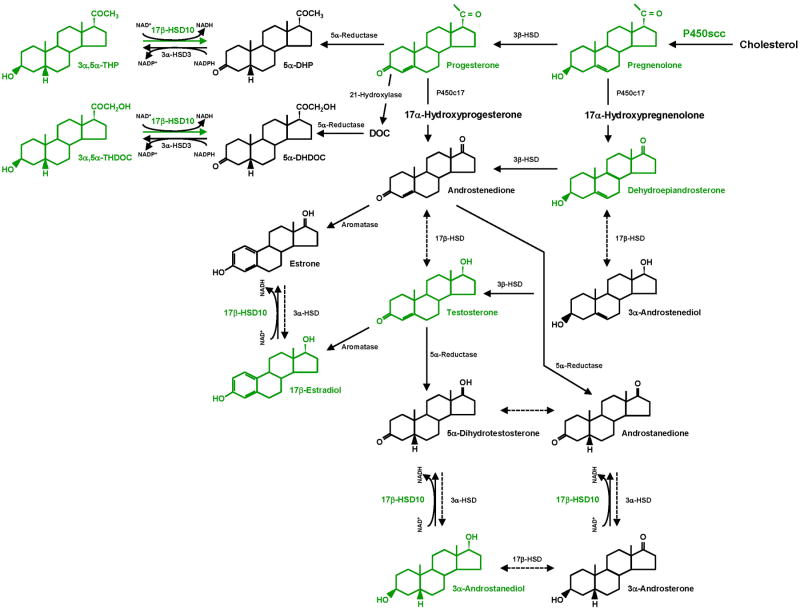
Neuroactive steroids are endogenous neuromodulators synthesised in the brain that rapidly alter neuronal excitability by binding to membrane receptors, in addition to the regulation of gene expression via intracellular steroid receptors. Neuroactive steroids induce potent anxiolytic, antidepressant, anticonvulsant, sedative, analgesic and amnesic effects, mainly through interaction with the GABAA receptor. They also exert neuroprotective, neurotrophic and antiapoptotic effects in several animal models of neurodegenerative diseases. Neuroactive steroids regulate many physiological functions, such as the stress response, puberty, the ovarian cycle, pregnancy and reward. Their levels are altered in several neuropsychiatric and neurological diseases and both preclinical and clinical studies emphasise a therapeutic potential of neuroactive steroids for these diseases, whereby symptomatology ameliorates upon restoration of neuroactive steroid concentrations. However, direct administration of neuroactive steroids has several challenges, including pharmacokinetics, low bioavailability, addiction potential, safety and tolerability, which limit its therapeutic use. Therefore, modulation of neurosteroidogenesis to restore the altered endogenous neuroactive steroid tone may represent a better therapeutic approach. This review summarises recent approaches that target the neuroactive steroid biosynthetic pathway at different levels aiming to promote neurosteroidogenesis. These include modulation of neurosteroidogenesis through ligands of the translocator protein 18 kDa and the pregnane xenobiotic receptor, as well as targeting of specific neurosteroidogenic enzymes such as 17β-hydroxysteroid dehydrogenase type 10 or P450 side chain cleavage. Enhanced neurosteroidogenesis through these targets may be beneficial not only for neurodegenerative diseases, such as Alzheimer's disease and age-related dementia, but also for neuropsychiatric diseases, including alcohol use disorders.

Keywords: 17β-hydroxysteroid dehydrogenase type 10; 3α,5α-THP; Alzheimer's disease; P450 side chain cleavage; alcoholism; neuroactive steroids; pregnane xenobiotic receptor; translocator protein 18 kDa.


Figures

Figure 1
Figure 1
Outline of neurosteroidogenesis. Neuroactive steroids and neurosteroidogenic enzymes that are potential key therapeutic targets are shown in green. The side chain of cholesterol is cleaved by P450scc as cholesterol is transported to the inner mitochondrial membrane and thus converted to pregnenolone. Soluble pregnenolone can enter into the endoplasmic reticulum unaided. 17β-HSD10 catalyzes the oxidation of neuroactive steroids in mitochondria with NAD+ as the coenzyme. This enzyme most effectively catalyzes the oxidation of 3α,5α-THP and 3α,5α-THDOC such that it is essential for the homeostasis of these neuroactive steroids, which was controlled by a dual enzyme molecular switch, composed of 17β-HSD10 and 3α-hydroxysteroid dehydrogenase type III (AKR1C2) localized in distinct subcellular compartments, mitochondria and ER, respectively (164, 168). The catalytic efficiencies (kcat/Km) of 17β-HSD10 are as high as 427 and 1,381 min-1 •mM-1 for the oxidation of 3α,5α-THP and 3α,5α-THDOC, respectively (163, 164). Abbreviations: 5α-DHP, 5α-dihydroprogesterone; DOC, deoxycorticosterone; 5α-DHDOC, 5α-dihydrodeoxycorticosterone; 3α,5α-THP, (3α,5α)-3-hydroxypregnan-20-one or allopregnanolone; 3α,5α-THDOC, (3α,5α)-3,21-dihydroxypregnan-20-one or allotetrahydrodeoxycorticosterone; HSD, hydroxysteroid dehydrogenase.
Figure 2
Figure 2
Figure depicts behaviour, pregnane steroid concentrations, and expression patterns in the hippocampus (top panel) and cortex (bottom panel) for 12 month old transgenic mice that co-overexpress mutant forms of amyloid precursor protein and presenilin 1 Δ exon 9 mutation (APPswe+PSEN1Δe9; a murine model of early-onset familial Alzheimer's disease- AD), compared to their age-matched wild-type controls (n = 4-6). For all measures, mice had data collected at 12 months of age, following 6 months of continuous progesterone (P4) administration via subcutaneously implanted pellets (25 mg, 90-day release at 6 months of age and then 9 months of age; purchased from Innovative Research of America). Behaviour: Performance in a memory task assessing the hippocampus (object placement) and cortex (object recognition) is depicted and based upon comparing the Alzheimer's disease mice to wild-type controls (methods and raw data published in (146)). Pregnane steroid concentrations: Levels of P4, dihydroprogesterone (DHP), and 3α,5α-THP (THP) were measured using radioimmunoassay of dissected out hippocampus and cortex (methods and raw data published in (146)). Protein expression: Expression patterns in hippocampus and cortex were determined by western blotting, of specific proteins (pregnane xenobiotic receptor (PXR), cytochrome P450-dependent side chain cleavage- P450scc, steroidogenic acute regulatory protein- StAR, 3β-hydroxysteroid dehydrogenase- 3β-HSD, and 5α-reductase- 5α-R). The mean of relative intensity (relative density of protein of interest to actin control) in hippocampus and cortex of Alzheimer's disease mice were compared to that determined in wild-type controls. A standard western blotting protocol (146) was employed to assess these factors in hippocampus and prefrontal cortex tissues (a description of tissue collection from animal subjects, brain storage, dissection and preparation is described in (146)). Protein concentration for each sample was determined with a Nanodrop spectrometer. Samples of equal protein concentrations were then prepared for loading on to NuPAGE Bis-Tris Mini Gels (4-12% SDS Polyacrylamide) by combining them with 2.5 μl of NuPAGE LDS (4×) sample buffer, 1 μl of NuPAGE Reducing Agent (10×), 6.5 μl of deionized water (Invitrogen). Electrophoresis was then conducted with running gels with 1× MOPS running buffer with one lane reserved for the protein ladder and one for the positive control (liver homogenate). Protein was then transferred to nitrocellulose using 1× NuPAGE Transfer buffer. The blots were blocked in 5% milk PBS-10% tween solution. All blots were probed with primary antibodies (Ab) at 4°C overnight. Primary (1°) Ab for PXR, P450, StAR, and 5α-reductase were purchased from Santa Cruz Biotechnology; 3β-HSD was received from Dr. Penning, and actin was purchased from Sigma. The secondary (2°) Ab was a Goat Anti-Mouse IG (H+L) Horseradish Peroxidase Conjugate (Bio-rad, Hercules, CA, USA). Concentrations of Ab were: PXR (1° Ab concentration 1:1000; 2° Ab concentration 1:2500); P450 (1° Ab concentration 1:2500; 2° Ab concentration 1:2500); StAR (1° Ab concentration 1:1000; 2° Ab concentration 1:2500); 3β-HSD (1° Ab concentration 1:2500; 2° Ab concentration 1:2500); 5α-reductase (1° Ab concentration 1:1500; 2° Ab concentration 1:2500), actin (1° Ab concentration 1:500; 2° Ab concentration 1:2500). Blots were probed with 2° Ab for 1 hour on a shaker at room temperature (Bio-rad). Results were visualized using DuoLuX Chemiluminescent/Fluorescent Substrate Kit for Peroxidase (Vector Laboratories), imaged on a ChemiDoc XRS (Bio-rad), and analyzed using ImageJ software. Expression was not detected (ND) for 3β-HSD in either group or for 5α-reductase in the wild-type controls, albeit Alzheimer's disease mice showed 2.0 relative intensity. Relative intensity values in the hippocampus (mean ± sem) for PXR (wildtype 0.4 ± 0.2; AD 1.0 ± 0.2), P450 (wildtype 0.3 ± 0.1; AD 1.4 ± 0.3), StAR (wildtype 0.6 ± 0.1; AD 1.0 ± 0.3), 3α-HSD (wildtype 0.4 ± 0.2; AD 0.8 ± 0.5) and 5α-reductase (wildtype 0.5 ± 0.2; AD 1.0 ± 0.7). Relative intensity values in the cortex (mean ± sem) for PXR (wildtype 2.2 ± 1.2; AD 1.0 ± 0.3), P450 (wildtype 1.7 ± 0.8; AD 0.6 ± 0.1), StAR (wildtype 1.4 ± 0.8; AD 0.9 ± 0.2), 3α-HSD (wildtype ND; AD ND) and 5α-reductase (wildtype ND; AD 2.0 ± 1.2).

References

    1. Paul SM, Purdy RH. Neuroactive steroids. FASEB J. 1992;6(6):2311–22. - PubMed
    1. Baulieu EE. Neurosteroids: a novel function of the brain. Psychoneuroendocrinology. 1998;23(8):963–87. - PubMed
    1. Charlier TD, Cornil CA, Patte-Mensah C, Meyer L, Mensah-Nyagan AG, Balthazart J. Local modulation of steroid action: rapid control of enzymatic activity. Front Neurosci. 2015;9:83. - PMC - PubMed
    1. Agis-Balboa RC, Pinna G, Zhubi A, Maloku E, Veldic M, Costa E, Guidotti A. Characterization of brain neurons that express enzymes mediating neurosteroid biosynthesis. Proc Natl Acad Sci U S A. 2006;103(39):14602–7. - PMC - PubMed
    1. Belelli D, Lambert JJ. Neurosteroids: endogenous regulators of the GABAA receptor. Nat Rev Neurosci. 2005;6(7):565–75. - PubMed

Publication types

MeSH terms

Associated data