. 2016 Jan 29;44(2):509-23.
doi: 10.1093/nar/gkv1470. Epub 2015 Dec 17.

Biogenesis and growth phase-dependent alteration of 5-methoxycarbonylmethoxyuridine in tRNA anticodons

Affiliations

Biogenesis and growth phase-dependent alteration of 5-methoxycarbonylmethoxyuridine in tRNA anticodons

Yusuke Sakai et al. Nucleic Acids Res. .

Abstract

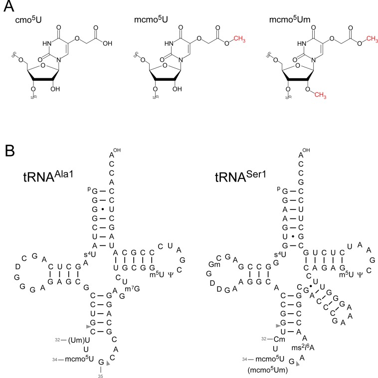
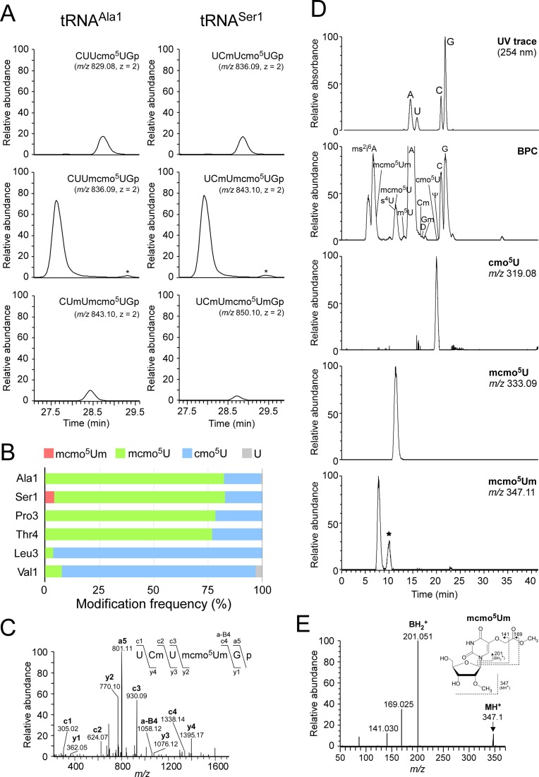
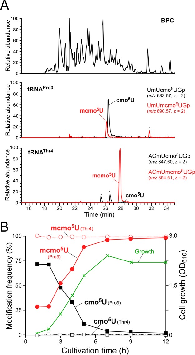
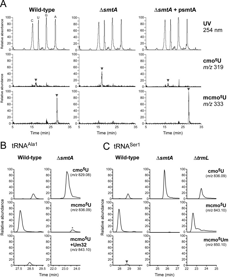
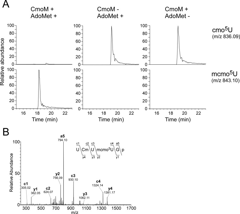
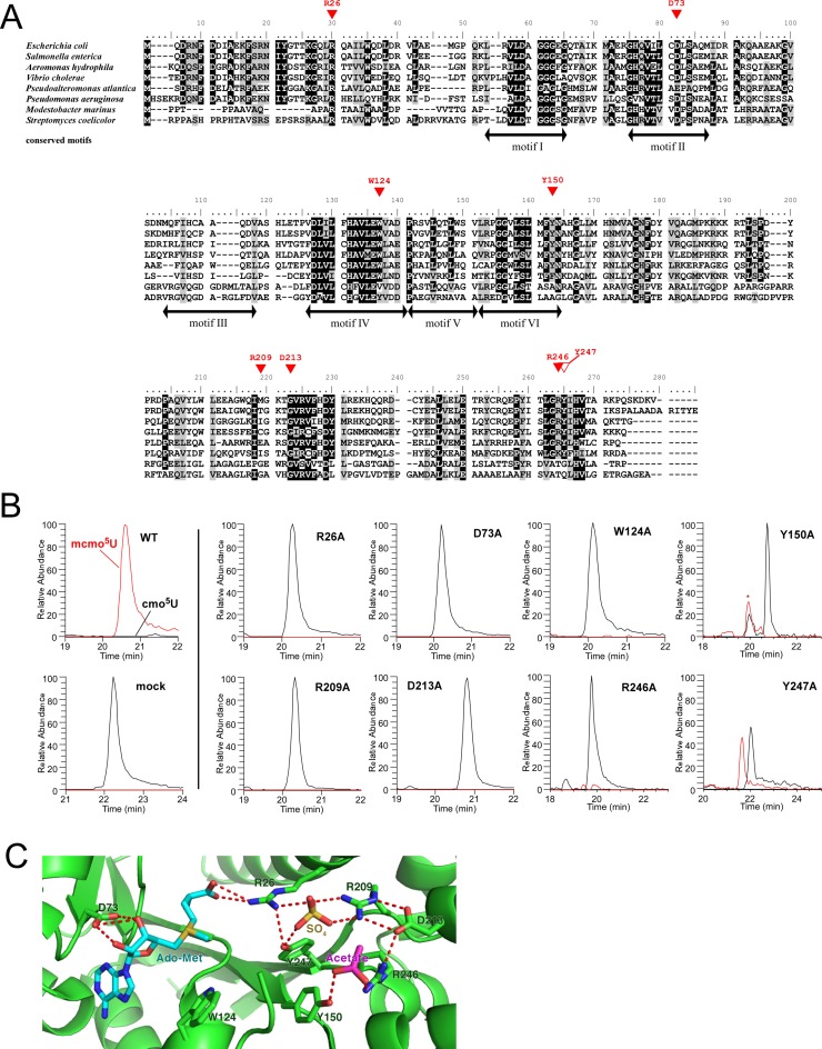
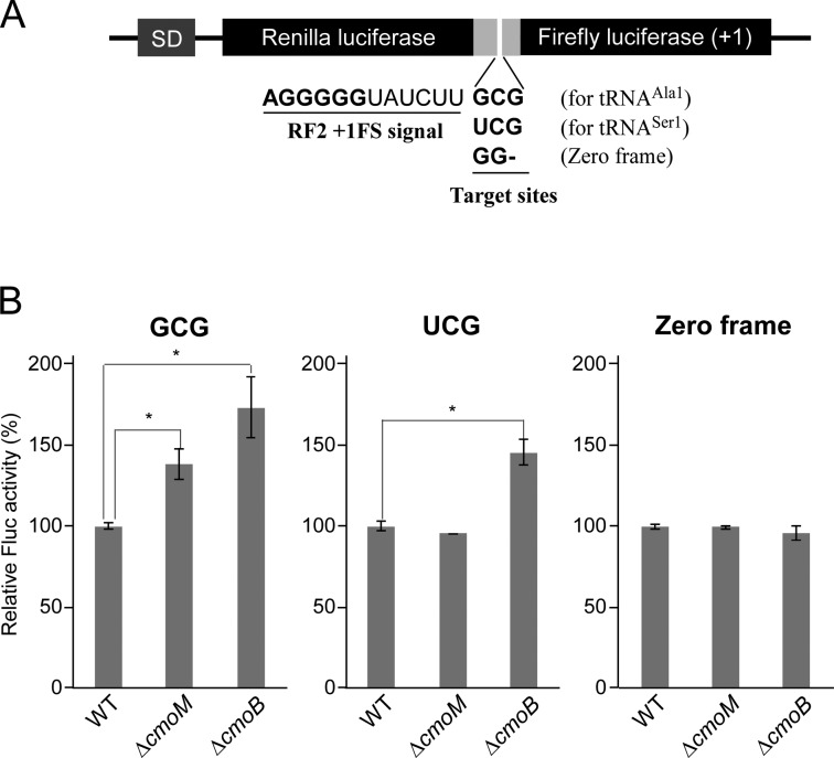
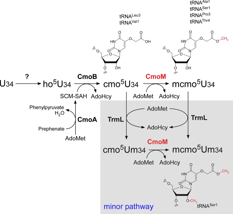
Post-transcriptional modifications at the anticodon first (wobble) position of tRNA play critical roles in precise decoding of genetic codes. 5-carboxymethoxyuridine (cmo(5)U) and its methyl ester derivative 5-methoxycarbonylmethoxyuridine (mcmo(5)U) are modified nucleosides found at the anticodon wobble position in several tRNAs from Gram-negative bacteria. cmo(5)U and mcmo(5)U facilitate non-Watson-Crick base pairing with guanosine and pyrimidines at the third positions of codons, thereby expanding decoding capabilities. By mass spectrometric analyses of individual tRNAs and a shotgun approach of total RNA from Escherichia coli, we identified mcmo(5)U as a major modification in tRNA(Ala1), tRNA(Ser1), tRNA(Pro3) and tRNA(Thr4); by contrast, cmo(5)U was present primarily in tRNA(Leu3) and tRNA(Val1). In addition, we discovered 5-methoxycarbonylmethoxy-2'-O-methyluridine (mcmo(5)Um) as a novel but minor modification in tRNA(Ser1). Terminal methylation frequency of mcmo(5)U in tRNA(Pro3) was low (≈30%) in the early log phase of cell growth, gradually increased as growth proceeded and reached nearly 100% in late log and stationary phases. We identified CmoM (previously known as SmtA), an AdoMet-dependent methyltransferase that methylates cmo(5)U to form mcmo(5)U. A luciferase reporter assay based on a +1 frameshift construct revealed that terminal methylation of mcmo(5)U contributes to the decoding ability of tRNA(Ala1).


Figures

Figure 1.
Figure 1.
5-carboxymethoxyuridine (cmo5U) and E. coli tRNAs. (A) Chemical structures of 5-carboxymethoxyuridine (cmo5U, left), 5-methoxycarbonylmethoxyuridine (mcmo5U, center) and 5-methoxycarbonylmethoxy-2′-O-methyluridine (mcmo5Um, right). (B) Secondary structures of E. coli tRNAAla1 (left) and tRNASer1 (right) with post-transcriptional modifications: 4-thiouridine (s4U), 2′-O-methylguanosine (Gm), dihydrouridine (D), 2′-O-methylcytidine (Cm), 2′-O-methyluridine (Um), 5-methoxycarbonylmethoxyuridine (mcmo5U), 5-methoxycarbonylmethoxy-2′-O-methyluridine (mcmo5Um), 2-methylthio-N6-isopentenyladenosine (ms2i6A), 7-methylguanosine (m7G), 5-methyluridine (m5U) and pseudouridine (Ψ). The position numbers of the residues (gray letters) are displayed according to the nucleotide numbering system (64). Pairs of gray triangles indicate the positions of cleavage by RNase T1 that generate RNA fragments containing the wobble positions. The sequence of E. coli tRNAAla1 is the same as that of tRNAAla1B (65).
Figure 2.
Figure 2.
Mass spectrometric analysis of individual tRNAs isolated from stationary-phase E. coli. (A) Mass chromatograms of RNase T1-digested fragments containing cmo5U and its derivatives from tRNAAla1 (left panels) and tRNASer1 (right panels) isolated from stationary-phase E. coli. Top and middle panels: extracted-ion chromatograms (XIC) for doubly-charged negative ions of cmo5U34-containing fragments (CUUcmo5UGp of tRNAAla1, MW 1660.18, m/z 829.08; UCmUcmo5UGp of tRNASer1, MW 1674.19, m/z 836.09) and mcmo5U34-containing fragments (CUUmcmo5UGp of tRNAAla1, MW 1674.19, m/z 836.09; UCmUmcmo5UGp of tRNASer1, MW 1688.21, m/z 843.10), respectively. Bottom left and bottom right panels: XICs for doubly-charged negative ions of Um32/mcmo5U34-containing fragment of tRNAAla1 (CUmUmcmo5UGp, MW 1688.21, m/z 843.10) and mcmo5Um34-containing fragment of tRNASer1 (UCmUmcmo5UmGp, MW 1702.22, m/z 850.10), respectively. The peaks marked with asterisks represent Um32/cmo5U-containing fragment (CUmUcmo5UGp, MW 1674.19, m/z 836.09) in tRNAAla1 and cmo5Um-containing fragment (UCmUcmo5UmGp, MW 1688.21, m/z 843.10) in tRNASer1. The RNA fragments containing unmodified C32 are also present in tRNASer1, but they are not described here due to high frequency of Cm32 in tRNASer1 isolated from stationary-phase E. coli. (B) Modification frequencies of cmo5U and its derivatives at position 34 in six species of tRNAs isolated from stationary-phase E. coli. Relative composition of each modification was calculated from the peak area ratio of mass chromatograms of RNase T1-digested fragments containing mcmo5Um34 (red), mcmo5U34 (green), cmo5U34 (blue) or U (gray). (C) Collision-induced dissociation (CID) spectrum of a fragment of E. coli tRNASer1 containing mcmo5Um34. The doubly-charged negative ion of the RNase T1-digested fragment containing mcmo5Um34 (m/z 850.10) was used as the precursor ion for CID. The product ions were assigned as described previously (66). Sequences of parent ion and assigned product ions are described on the upper left side of this panel. (D) Nucleoside analysis of E. coli tRNASer1. Top panel: UV chromatogram at 254 nm of the four major nucleosides (C, U, G and A). Second panel: mass chromatograms of the protonated nucleosides (MH+) on the base peak chromatogram (BPC). Third to bottom panels: XICs for cmo5U (m/z 319.08), mcmo5U (m/z 333.09) and mcmo5Um (m/z 347.11), respectively. The peak marked with an asterisk represents unspecific peak. (E) CID spectrum of mcmo5Um nucleoside. The protonated mcmo5Um (MH+, m/z 347.11) was used as the precursor ion for CID. The N-glycoside bond cleaved to generate the base-related ion (BH2+) and other product ions are assigned on the chemical structure.
Figure 3.
Figure 3.
Shotgun analysis of tRNA fragments and growth phase-dependent alteration of mcmo5U. (A) Shotgun analysis of total tRNA digested by RNase T1. Top panel: base peak chromatogram (BPC) of RNase T1-digested total tRNA isolated from early log phase (2 h) E. coli. Second panel: XICs for doubly-charged negative ions of the cmo5U34-containing fragment (UmUcmo5UGp, m/z 683.57) and the mcmo5U34-containing fragment (UmUmcmo5UGp, m/z 690.57) from tRNAPro3. Bottom panel: XICs for doubly-charged negative ions of the cmo5U34-containing fragment (ACmUcmo5UGp, m/z 847.60) and the mcmo5U34-containing fragment (ACmUmcmo5UGp, m/z 854.61) from tRNAThr4. (B) mcmo5U level is altered by growth phase. tRNAPro3 (filled circle and rectangle) or tRNAThr4 (open circle and rectangle) were calculated from the peak area ratio of XICs for the fragments containing mcmo5U34 and cmo5U34 at different cultivation times. Cell growth (green line) was monitored by absorbance at 610 nm.
Figure 4.
Figure 4.
Reverse genetic approach identified a gene responsible for terminal methylation of mcmo5U. (A) Nucleoside analyzes by LC/MS using reverse phase chromatography of total RNA from wild-type (left panels), ΔsmtA (middle panels) and ΔsmtA rescued with psmtA (right panels). Top panels: UV trace at 254 nm. Second and bottom panels: XICs for cmo5U (m/z 319) and mcmo5U (m/z 333), respectively. Intensity of each peak was normalized to that of cyclic t6A (m/z 395). (B) Mass chromatograms of RNase T1-digested fragments containing cmo5U and its derivatives from tRNAAla1 isolated from wild-type (left panels) and ΔsmtA (right panels) strains. Top, middle and bottom panels: XICs for doubly-charged negative ions of the cmo5U34-containing fragments (CUUcmo5UGp, m/z 829.08), the mcmo5U34-containing fragments (CUUmcmo5UGp, m/z 836.09) and the Um32/mcmo5U34-containing fragment (CUmUmcmo5UGp, m/z 843.10), respectively. The peak marked with an asterisk represent the Um32/cmo5U-containing fragment (CUmUcmo5UGp, m/z 836.09). (C) Mass chromatograms of RNase T1-digested fragments containing cmo5U and its derivatives from tRNASer1 isolated from wild-type (left panels), ΔsmtA (middle panels) and ΔtrmL (right panels) strains. Top, middle and bottom panels: XICs for doubly-charged negative ions of the cmo5U34-containing fragments (UCmUcmo5UGp, m/z 836.09), the mcmo5U34-containing fragments (UCmUmcmo5UGp, m/z 843.10) and the mcmo5Um34-containing fragment (UCmUmcmo5UmGp, m/z 850.10), respectively.
Figure 5.
Figure 5.
In vitro reconstitution of cmo5U methylation by recombinant CmoM. (A) E. coli tRNASer1 bearing cmo5U isolated from the ΔcmoM strain was incubated in the presence or absence of recombinant CmoM with or without AdoMet. Top and bottom panels: XICs for doubly-charged negative ions of the cmo5U34-containing fragments (UCmUcmo5UGp, m/z 836.09) and the mcmo5U34-containing fragments (UCmUmcmo5UGp, m/z 843.10), respectively. (B) A CID spectrum of RNase T1-digested fragment of E. coli tRNASer1 incubated in the presence of recombinant CmoM with AdoMet. The doubly-charged negative ion of the mcmo5U34-containing fragment (UCmUmcmo5UGp, m/z 843.10) was used as the precursor ion for CID. The product ions were assigned according to the literature (66). Sequences of parent ion and assigned product ions are described upper left side in this panel.
Figure 6.
Figure 6.
Characterization of CmoM. (A) Sequence alignment of CmoM homologs from six γ-proteobacteria, Escherichia coli (NP_415441.1), Salmonella enterica (NP_455477.1), Aeromonas hydrophila (YP_856903.1), Vibrio cholerae (NP_231353.1), Pseudoalteromonas atlantica (ABG40695.1), Pseudomonas aeruginosa (NP_253478.1), and two Actinobacteria, Modestobacter marinus (WP_014741474.1), Streptomyces coelicolor (WP_011028138.1). Identical or similar residues are shaded in black or gray, respectively. Red triangles indicate residues that are essential (filled) or non-essential (open) for generic complementation. Motifs I to VI are conserved in Class I AdoMet-dependent methyltransferases. (B) XICs for doubly-charged negative ions of the cmo5U34-containing fragment (black lines, UmUcmo5UGp, m/z 683.57) and the mcmo5U34-containing fragment (red lines, UmUmcmo5UGp, m/z 690.57) from tRNAPro3 in the ΔcmoM strain rescued by plasmid-encoded wild-type cmoM or its mutant derivatives. The peak marked with an asterisk represents unspecific peak. (C) Close-up view of the AdoMet-binding site in the crystal structure of E. coli CmoM (PDB ID: 4HTF) containing ligands, AdoMet, acetate and sulfate. Predicted hydrogen bonds between ligands and CmoM are indicated by red dotted lines.
Figure 7.
Figure 7.
Terminal methylation of mcmo5U contributes to GCG decoding. (A) Schematic depiction of the dual-luciferase reporter constructs based on the RF2 recoding system. SD, Shine-Dalgarno sequence. Renilla and firefly luciferases were fused with a linker containing the +1 frameshift signal of the RF2 recoding site. The frameshift target site was replaced with a GCG codon for tRNAAla1, a UCG codon for tRNASer1or GG for zero frame (used as a control). (B) Relative pausing activity at the frameshift site with GCG (left), UCG (middle) or zero frame (right) was calculated based on relative Fluc activity normalized to Rluc activity in wild-type, ΔcmoM and ΔcmoB strains. Data are presented as means ± SD (n = 4). *, P < 0.01 versus control (Student's t-test).
Figure 8.
Figure 8.
Biosynthesis of mcmo5U. In E. coli, U34 of six tRNAs responsible for decoding NCN codons is modified to ho5U34 by an unknown pathway, and subsequently modified to cmo5U in a reaction catalyzed by CmoM. CmoB uses SCM-SAH as a substrate and transfers its carboxymethyl group to ho5U34 on tRNAs. SCM-SAH is synthesized from prephenate and AdoMet catalyzed by CmoA. Four tRNAs (Ala1, Ser1, Pro3 and Thr4) are further modified to mcmo5U by CmoM, using AdoMet as a substrate. mcmo5U frequency is altered by growth phase only in tRNAPro3. In a minor pathway, mcmo5U34 in tRNASer1 is further methylated by TrmL to yield mcmo5Um34. Alternatively, cmo5U34 could be first converted to cmo5Um34, then to mcmo5Um34.

References

    1. Machnicka M.A., Milanowska K., Osman Oglou O., Purta E., Kurkowska M., Olchowik A., Januszewski W., Kalinowski S., Dunin-Horkawicz S., Rother K.M., et al. MODOMICS: a database of RNA modification pathways–2013 update. Nucleic Acids Res. 2013;41:D262–D267. - PMC - PubMed
    1. Yokoyama S., Nishimura S. In: tRNA: Structure, Biosynthesis, and Function. Soll DRUL, editor. Washington, D.C.: American Society for Microbiology; 1995. pp. 207–224.
    1. Bjork G. In: tRNA: Structure, Biosynthesis, and Function. Soll DRUL, editor. Washington, D.C.: American Society for Microbiology; 1995. pp. 165–205.
    1. Suzuki T. In: Fine-Tuning of RNA Functions by Modification and Editing. Grosjean H, editor. Vol. 12. Springer-Verlag Berlin and Heidelberg: GmbH & Co. KG; 2005. pp. 23–69.
    1. Crick F.H. Codon–anticodon pairing: the wobble hypothesis. J. Mol. Biol. 1966;19:548–555. - PubMed

Publication types

MeSH terms