. 2017 Mar;66(3):454-463.
doi: 10.1136/gutjnl-2015-309453. Epub 2015 Dec 17.

T cell neoepitope discovery in colorectal cancer by high throughput profiling of somatic mutations in expressed genes

Affiliations

T cell neoepitope discovery in colorectal cancer by high throughput profiling of somatic mutations in expressed genes

Daniele Mennonna et al. Gut. 2017 Mar.

Abstract

Objective: Patient-specific (unique) tumour antigens, encoded by somatically mutated cancer genes, generate neoepitopes that are implicated in the induction of tumour-controlling T cell responses. Recent advancements in massive DNA sequencing combined with robust T cell epitope predictions have allowed their systematic identification in several malignancies.

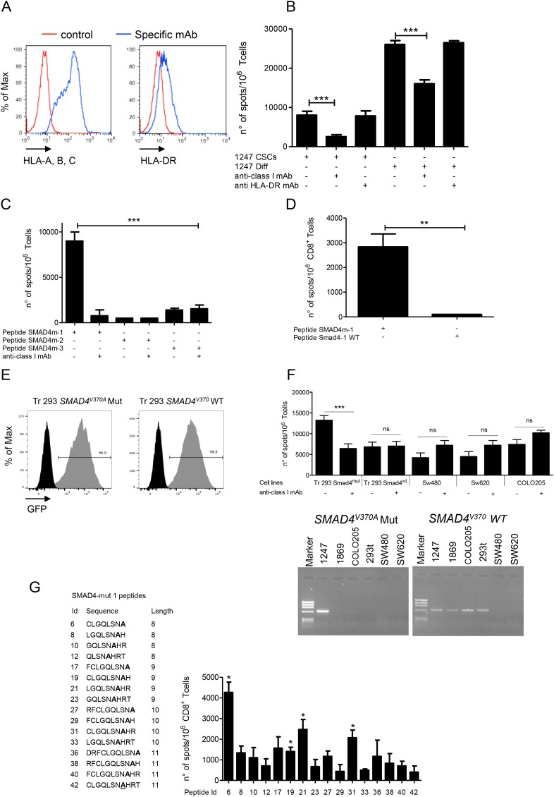
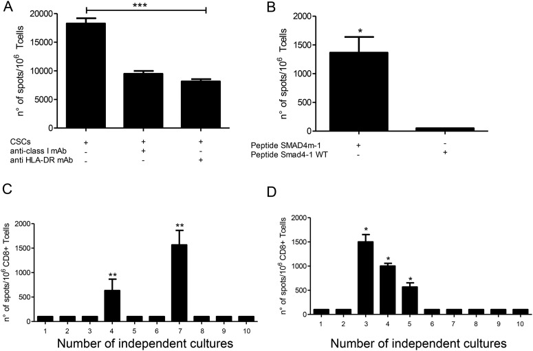
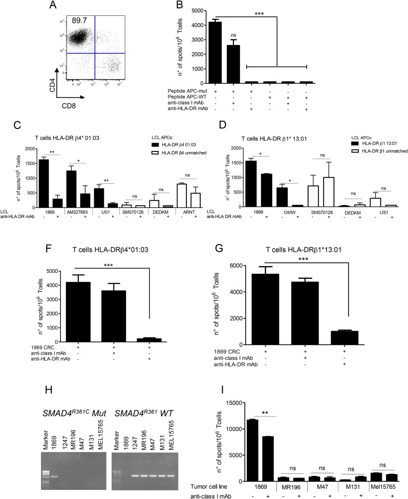
Design: We undertook the identification of unique neoepitopes in colorectal cancers (CRCs) by using high-throughput sequencing of cDNAs expressed by standard cancer cell cultures, and by related cancer stem/initiating cells (CSCs) cultures, coupled with a reverse immunology approach not requiring human leukocyte antigen (HLA) allele-specific epitope predictions.

Results: Several unique mutated antigens of CRC, shared by standard cancer and related CSC cultures, were identified by this strategy. CD8+ and CD4+ T cells, either autologous to the patient or derived from HLA-matched healthy donors, were readily expanded in vitro by peptides spanning different cancer mutations and specifically recognised differentiated cancer cells and CSC cultures, expressing the mutations. Neoepitope-specific CD8+ T cell frequency was also increased in a patient, compared with healthy donors, supporting the occurrence of clonal expansion in vivo.

Conclusions: These results provide a proof-of-concept approach for the identification of unique neoepitopes that are immunogenic in patients with CRC and can also target T cells against the most aggressive CSC component.

Keywords: ANTIGENS; CANCER IMMUNOBIOLOGY; COLORECTAL CANCER; GENE MUTATION; IMMUNE RESPONSE.


Conflict of interest statement

Competing interests: None declared.

Figures

Figure 1
Figure 1
Patient T cells recognise a SMAD4V370A-containing neoepitope presented by autologous cancer cells. (A) HLA class I and HLA-DR expression in 1247 cancer cells following 48 h IFNγ induction. (B–D) IFNγ ELISPOT of CD8+ T cells from 1247 patient, stimulated with a peptide pool encompassing the SMAD4V370A mutation, assayed for the recognition of: 1247 cancer stem/initiating cells (CSCs) and differentiated colorectal cancer (CRC) cells (B), of SMAD4 mutated peptides presented by HLA-A*0201+ T2 cells (C), of SMAD4m-1 and SMAD41 WT peptides presented by T2 cells (D), ±anti-class I or HLA-DR mAbs. (E) Percentage of GFP expression by sorted HEK293t cells transduced with retroviral vectors encoding the 1247 SMAD4V37A mutated or the corresponding SMAD4V37 WT minigenes; (F) Upper panel. SMAD4V37A-specific CD8+ T cells assayed by IFNγ ELISPOT for the recognition of HEK293t cells transduced with the SMAD4 minigenes, or of three HLA-A*02:01+ CRC cell lines negative for the SMAD4V37A mutation, ±anti-class I mAb. Lower panel. PCR typing for the expression of the SMAD4V37A mutation, or the corresponding SMAD4V37 WT sequence in the CRC cell lines shown in the upper panel, and in untransduced HEK293t (293t) cells. The 1247 and 1869 CRC cell lines are positive and negative controls for the PCR, respectively. (G) SMAD4m-1 peptide-stimulated CD8+ T cells assayed by IFNγ ELISPOT (right panel) for the specific recognition of the peptide epitopes of different lengths (left panel), presented by T2 cells. All IFNγ ELISPOT data are triplicate mean±SD, subtracted of the background spots produced by T cells alone, and are representative of three independent experiments performed with independently induced CD8+ T cell lines. Only the experiment in panel F was performed twice with the same CD8+ T cell line. *p≤0.05; **p≤0.01; ***p≤0.001; ns, non-statistically significant.
Figure 2
Figure 2
The SMAD4V370A mutation of 1247 colorectal cancer (CRC) is spontaneously immunogenic for autologous CD8+ T cells. (A) Mixed lymphocyte-tumour cell culture (MLTC) from patient 1247, induced by autologous cancer stem/initiating cell (CSC) cultures, assayed by IFNγ ELISPOT for the recognition of the autologous CSCs, ±anticlass I mAb or anti-HLA-DR mAb. (B) CD8+ T cells, enriched from the previous MLTC, assayed by IFNγ ELISPOT for the recognition of the SMAD4m-1 or SMAD4-1 WT peptides presented by T2 cells; (C) Primary CD8+ T cells purified from patient 1247, stimulated twice with the SMAD4-1 peptide in the presence of autologous PBMCs in two series of 10 wells, each containing 5×10⁴ (left panel) or 105 (right panel) cells/well for a total of 1.5×10⁶ precursors, and assayed by IFNγ ELISPOT for the recognition of the 1247 CRC cells. IFNγ ELISPOT data are represented as triplicate mean±SD, subtracted of the background spots produced by T cells alone, and are representative of three (A) and two (B) independent experiments performed. *p≤0.05; **p≤0.01; ***p≤0.001.
Figure 3
Figure 3
The 1869 colorectal cancer (CRC) presents neoepitopes from mutated genes to allogeneic CD4+ and CD8+ T cells. (A) CD4+ phenotype of a T cell line from a HLA-DR*β4 01:03 healthy donor (HD) expanded with the 30 aa long antigen presenting cell (APC)E1317KfsX4 synthetic peptide. (B) T cells from the HLA-DR*β4 01:03 HD assayed by IFNγ ELISPOT for the recognition of 1869 B lymphoblastoid cell lines (LCLs) loaded with the APC E1317KfsX4 or the WT peptide, ±anti-HLA-DR mAb. Similar results were obtained with CD4+ T cell lines elicited by the APC E1317KfsX4 peptide from HLA-DR*β1 13:01 HDs. (C–D) APCE1317KfsX4-specific CD4+ T cells from either HLA-DR*β4 01:03 or HLA-DR*β113:01 donor assayed by IFNγ ELISPOT for the recognition of LCL cells homozygous for either HLA-DR*β4 01:03 (C) or HLA-DR*β113:01 (D), or negative for these alleles, loaded with the APCE1317KfsX4 peptide±anti-HLA-DR mAb. (F–G) APCE1317KfsX4-specific CD4+ T cells induced from HLA-DR*β4 01:03- or HLA-DR*β1 13:01-matched HDs, assayed by IFNγ ELISPOT for the recognition of 1869 CRC cells, ±anti-HLA-DR mAbs. (H) PCR typing for the expression of the SMAD4R361C mutation, or the corresponding SMAD4V37 WT sequence, in the HLA-B*35:01+ cancer cell lines used for the recognition assay. The 1869 and 1247 cell lines are positive and negative control of the PCR, respectively. (I) CD8+ T cells elicited from HLA-B*35:01 HD with the peptide pool encompassing the SMAD4R361C mutation assayed by IFNγ ELISPOT for the recognition of 1869 CRC cells, or of HLA-B*35:01+ and SMAD4R361C-negative kidney cancer cell line MR196 and melanoma cell lines M47, M131 and Mel15765,±anti-class I mAb. IFNγ ELISPOT data are represented as triplicate mean±SD, subtracted of the background spots produced by T cells alone, and are representative of three independent experiments. *p≤0.05; **p≤0.01; ***p≤0.001; ns, non-statistically significant.

References

    1. Rosenberg SA, Yang JC, Sherry RM, et al. . Durable complete responses in heavily pretreated patients with metastatic melanoma using T-cell transfer immunotherapy. Clin Cancer Res 2011;17:4550–7. 10.1158/1078-0432.CCR-11-0116 - DOI - PMC - PubMed
    1. Hodi FS, O'Day SJ, McDermott DF, et al. . Improved survival with ipilimumab in patients with metastatic melanoma. N Engl J Med 2010;363:711–23. 10.1056/NEJMoa1003466 - DOI - PMC - PubMed
    1. Topalian SL, Hodi FS, Brahmer JR, et al. . Safety, activity, and immune correlates of anti-PD-1 antibody in cancer. N Engl J Med 2012;366: 2443–54. 10.1056/NEJMoa1200690 - DOI - PMC - PubMed
    1. Brahmer JR, Tykodi SS, Chow LQ, et al. . Safety and activity of anti-PD-L1 antibody in patients with advanced cancer. N Engl J Med 2012;366:2455–65. 10.1056/NEJMoa1200694 - DOI - PMC - PubMed
    1. Coulie PG, Van den Eynde BJ, van der Bruggen P, et al. . Tumour antigens recognized by T lymphocytes: at the core of cancer immunotherapy. Nature reviews Cancer 2014;14:135–46. 10.1038/nrc3670 - DOI - PubMed

Publication types

MeSH terms