

Assessment of Venous Thrombosis in Animal Models
- PMID: 26681755
- PMCID: PMC4805178
- DOI: 10.1161/ATVBAHA.115.306255
Assessment of Venous Thrombosis in Animal Models
Abstract
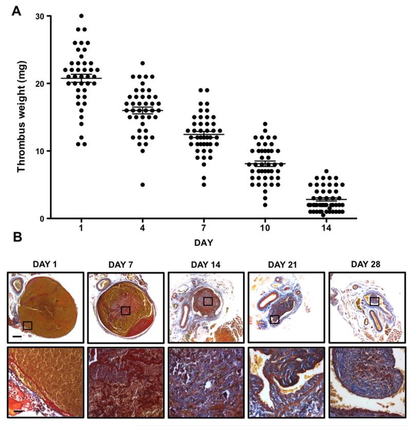
Deep vein thrombosis and common complications, including pulmonary embolism and post-thrombotic syndrome, represent a major source of morbidity and mortality worldwide. Experimental models of venous thrombosis have provided considerable insight into the cellular and molecular mechanisms that regulate thrombus formation and subsequent resolution. Here, we critically appraise the ex vivo and in vivo techniques used to assess venous thrombosis in these models. Particular attention is paid to imaging modalities, including magnetic resonance imaging, micro-computed tomography, and high-frequency ultrasound that facilitate longitudinal assessment of thrombus size and composition.
Keywords: histological techniques; models, animal; molecular imaging; venous thrombosis.
© 2015 American Heart Association, Inc.
Figures


References
-
- Cohen AT, Agnelli G, Anderson FA, Arcelus JI, Bergqvist D, Brecht JG, Greer IA, Heit JA, Hutchinson JL, Kakkar AK, Mottier D, Oger E, Samama MM, Spannagl M. Venous thromboembolism (vte) in europe. The number of vte events and associated morbidity and mortality. Thromb Haemost. 2007;98:756–764. - PubMed
-
- Hunt BJ. Awareness and politics of venous thromboembolism in the united kingdom. Arterioscler Thromb Vasc Biol. 2008;28:398–399. - PubMed
-
- Prandoni P, Lensing AW, Cogo A, Cuppini S, Villalta S, Carta M, Cattelan AM, Polistena P, Bernardi E, Prins MH. The long-term clinical course of acute deep venous thrombosis. Ann Intern Med. 1996;125:1–7. - PubMed
-
- Singh I, Burnand KG, Collins M, Luttun A, Collen D, Boelhouwer B, Smith A. Failure of thrombus to resolve in urokinase-type plasminogen activator gene-knockout mice: Rescue by normal bone marrow-derived cells. Circulation. 2003;107:869–875. - PubMed
-
- McGuinness CL, Humphries J, Waltham M, Burnand KG, Collins M, Smith A. Recruitment of labelled monocytes by experimental venous thrombi. Thromb Haemost. 2001;85:1018–1024. - PubMed
Publication types
MeSH terms
Substances
Grants and funding
LinkOut - more resources
Full Text Sources
Other Literature Sources
Medical
