. 2015:2015:730121.
doi: 10.1155/2015/730121. Epub 2015 Nov 22.

Resveratrol Protects PC12 Cell against 6-OHDA Damage via CXCR4 Signaling Pathway

Affiliations

Resveratrol Protects PC12 Cell against 6-OHDA Damage via CXCR4 Signaling Pathway

Jing Zhang et al. Evid Based Complement Alternat Med. 2015.

Abstract

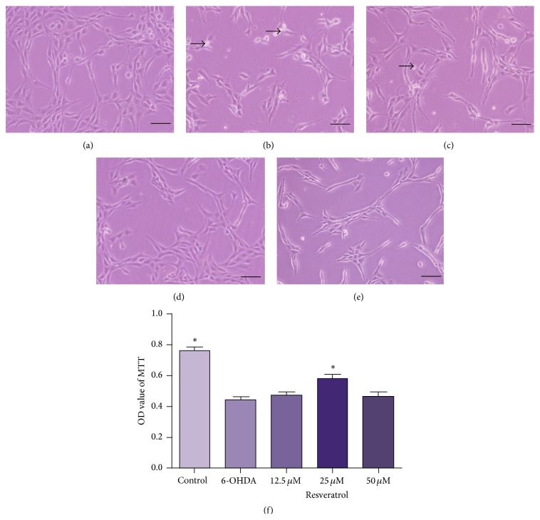
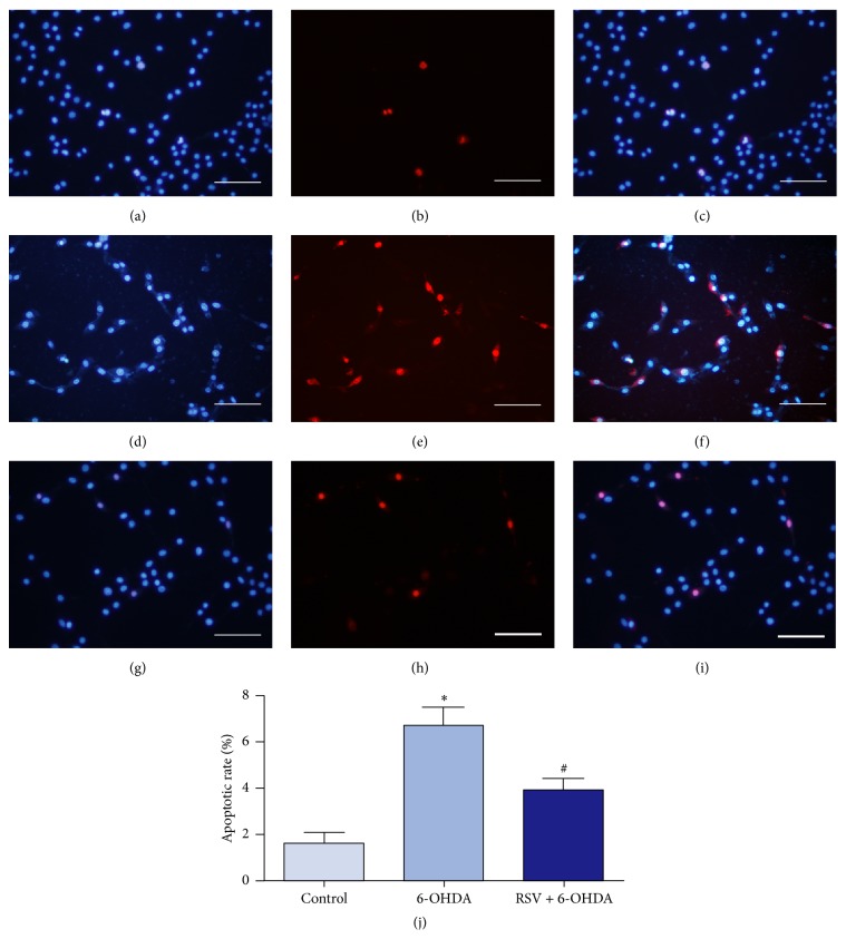
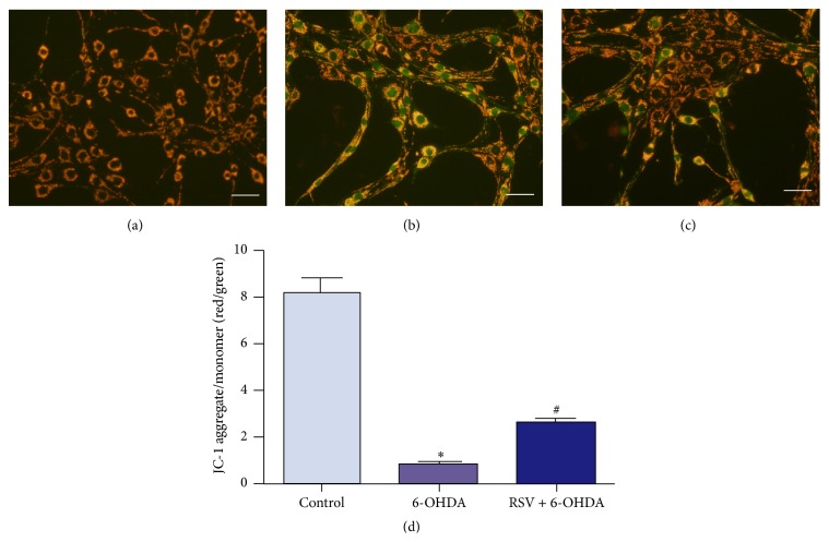
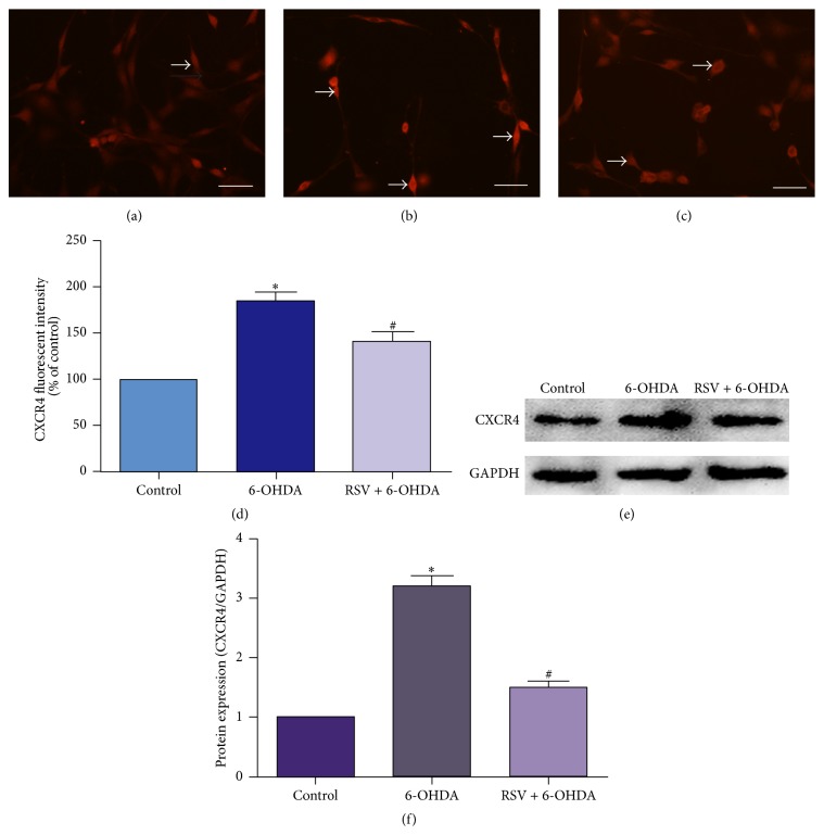
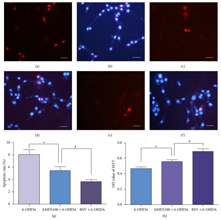
Resveratrol, herbal nonflavonoid polyphenolic compound naturally derived from grapes, has long been acknowledged to possess extensive biological and pharmacological properties including antioxidant and anti-inflammatory ones and may exert a neuroprotective effect on neuronal damage in neurodegenerative diseases. However, the underlying molecular mechanisms remain undefined. In the present study, we intended to investigate the neuroprotective effects of resveratrol against 6-OHDA-induced neurotoxicity of PC12 cells and further explore the possible mechanisms involved. For this purpose, PC12 cells were exposed to 6-OHDA in the presence of resveratrol (0, 12.5, 25, and 50 μM). The results showed that resveratrol increased cell viability, alleviated the MMP reduction, and reduced the number of apoptotic cells as measured by MTT assay, JC-1 staining, and Hoechst/PI double staining (all p < 0.01). Immunofluorescent staining and Western blotting revealed that resveratrol averts 6-OHDA induced CXCR4 upregulation (p < 0.01). Our results demonstrated that resveratrol could effectively protect PC12 cells from 6-OHDA-induced oxidative stress and apoptosis via CXCR4 signaling pathway.


Figures

Figure 1
Figure 1
6-OHDA induced damage to the cultured PC12 cells. (a–e) The photomicrographs of PC12 cells exposed to 6-OHDA of different concentration for 24 h. (a) PC12 cells in routine culture medium grew well with long neurites. (b) When exposed to 25 μM 6-OHDA, cell morphology had little changes. (c) When treated with 50 μM 6-OHDA, the neurites started to retract, and the neural network was collapsed in a certain degree with cell debris that appeared. (d and e) As the concentration of 6-OHDA increased to 100 and 150 μM, the cells were strongly insulted and severe cell loss could be observed. There was a mass of cell debris that could be found in the medium. Arrows indicated cell debris. (f) MTT assay also demonstrated that cell viability decreased gradually with the concentration of 6-OHDA increased. OD values were presented as means ± SD from five independent experiments. p < 0.01 versus control group. Scale bars: (a–e) 100 μm.
Figure 2
Figure 2
Protection of resveratrol on PC12 cells against 6-OHDA. (a) PC12 cells of normal control group grew in good condition and exhibited long neurites. (b) In 6-OHDA injury group, neurites were short and few with the neural network collapsed. (c–e) The cells were preincubated for 2 h with resveratrol at 12.5 (c), 25 (d), and 50 μM (e) before 50 μM 6-OHDA exposure, respectively. In 12.5 μM resveratrol group, the protective effect was weak with swollen cells and cell debris observed (c). 25 μM resveratrol promoted neurite growth, and only little cell debris could be found (d). However, in 50 μM resveratrol group, the alleviative effect was not obvious, and some cells swelled and turned into a round shape (e). Arrows (b and c) indicated cell debris. Cell viability was further measured by MTT assays (f). OD values were presented as means ± SD from five independent experiments. p < 0.01 versus 6-OHDA injury group. Scale bars: (a–d) 100 μm.
Figure 3
Figure 3
Resveratrol prevents 6-OHDA-induced cell apoptosis. PC12 cells were exposed to 6-OHDA with or without resveratrol for 24 h and double stained with Hoechst 33342 (blue) and PI (red) to determine cell apoptosis. In the control group, PC12 cells were cultured in DMEM, and most of the cells displayed normal nuclear morphology with uniform blue nuclei (a–c). In 6-OHDA injury group, the numbers of cells with bright red nuclei were obviously increased (d–f). And only few apoptotic cells could be observed when preincubated with 25 μM resveratrol (g–i). (j) Statistical analysis showed that, compared with the control group, PC12 cells treated with 6-OHDA exhibited greater apoptotic rate ( p < 0.01, n = 5), while when preincubated with 25 μM resveratrol, the apoptotic rate was largely decreased (# p < 0.01, n = 5). Scale bars: (a–i) 100 μm.
Figure 4
Figure 4
Effects of 6-OHDA and resveratrol on MMP of PC12 cells. (a–c) The fluorescent images of PC12 cells stained with JC-1 dye and captured by a Zeiss 780 laser confocal microscope. In the normal control group, cells exhibited clearly orange red (a), while, in 6-OHDA injury group, quite amounts of cells had high level of green fluorescent intensity, with lower level of red fluorescence (b). When preincubated with resveratrol, the green fluorescent intensity was effectively decreased meanwhile red fluorescence was increased (c). (d) Quantification of fluorescent intensity of PC12 cells in different group. Statistical analysis showed that the red/green fluorescence intensity ratio of PC12 cells treated with 6-OHDA was obviously decreased, which could be prevented by 25 μM resveratrol. Ratio of red/green fluorescence was presented as means ± SD from five independent experiments. p < 0.01 versus control group, # p < 0.01 versus 6-OHDA injury group. Scale bars: (a–c) 50 μm.
Figure 5
Figure 5
Resveratrol downregulates the expression of CXCR4 in PC12 cells treated with 6-OHDA. Immunofluorescent staining (a–d) and Western blotting (e, f) were used to determine the expression of CXCR4. (a) In normal control group, the intensity of the staining was weak. After being treated with 6-OHDA, the fluorescent intensity was increased (b). In the presence of resveratrol, this increase was effectively decreased (c). Arrows (a, b, and c) indicated the positive fluorescent staining. (d) Quantification of fluorescent intensity of PC12 cells in different group. In Western blotting, the level of CXCR4 protein was significantly increased in cells treated with 6-OHDA and preincubation with resveratrol could decrease the upregulated protein levels of CXCR4 (e and f). Values of fluorescent intensity were presented as means ± SD from five independent experiments. p < 0.01 versus control group, # p < 0.01 versus 6-OHDA injury group. Scale bars: (a–c) 100 μm.
Figure 6
Figure 6
CXCR4 inhibitor AMD3100 protected PC12 cells from 6-OHDA induced damage. To further investigate the role of CXCR4 in 6-OHDA induced neurotoxicity, AMD3100 was added into the culture medium before 6-OHDA treated. Cell apoptosis was detected by Hoechst 33342 (blue) and PI (red) double staining (a–f) and observed under Zeiss LSM 780 laser confocal microscope. The number of apoptotic cells in AMD3100 group (c, d) was less than that in 6-OHDA group (a, b) but still more than that in resveratrol protection group (e, f). Cell apoptosis analysis (g) and MTT assay (h) demonstrated that AMD3100 could protect PC12 cells from injury induced by 6-OHDA. Data were presented as means ± SD. p < 0.05 versus 6-OHDA injury group, # p < 0.05 versus resveratrol protection group (n = 3). Scale bars: (a–f) 50 μm.

References

    1. Braak H., Del Tredici K., Rüb U., de Vos R. A. I., Jansen Steur E. N. H., Braak E. Staging of brain pathology related to sporadic Parkinson's disease. Neurobiology of Aging. 2003;24(2):197–211. doi: 10.1016/s0197-4580(02)00065-9. - DOI - PubMed
    1. Jankovic J. Parkinson's disease: clinical features and diagnosis. Journal of Neurology, Neurosurgery and Psychiatry. 2008;79(4):368–376. doi: 10.1136/jnnp.2007.131045. - DOI - PubMed
    1. Ward C. D., Hess W. A., Calne D. B. Olfactory impairment in Parkinson's disease. Neurology. 1983;33(7):943–946. doi: 10.1212/WNL.33.7.943. - DOI - PubMed
    1. Aarsland D., Andersen K., Larsen J. P., et al. The rate of cognitive decline in Parkinson disease. Archives of Neurology. 2004;61(12):1906–1911. doi: 10.1001/archneur.61.12.1906. - DOI - PubMed
    1. Bonito-Oliva A., Masini D., Fisone G. A mouse model of non-motor symptoms in Parkinson's disease: focus on pharmacological interventions targeting affective dysfunctions. Frontiers in Behavioral Neuroscience. 2014;8, article 290 doi: 10.3389/fnbeh.2014.00290. - DOI - PMC - PubMed

LinkOut - more resources