Preoperative portal vein embolization in liver cancer: indications, techniques and outcomes
- PMID: 26682142
- PMCID: PMC4671969
- DOI: 10.3978/j.issn.2223-4292.2015.10.04
Preoperative portal vein embolization in liver cancer: indications, techniques and outcomes
Erratum in
-
Erratum to preoperative portal vein embolization in liver cancer: indications, techniques and outcomes.Quant Imaging Med Surg. 2016 Oct;6(5):619-620. doi: 10.21037/qims.2016.10.13. Quant Imaging Med Surg. 2016. PMID: 27942484 Free PMC article.
Abstract
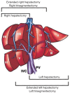
Postoperative liver failure is a severe complication of major hepatectomies, in particular in patients with a chronic underlying liver disease. Portal vein embolization (PVE) is an approach that is gaining increasing acceptance in the preoperative treatment of selected patients prior to major hepatic resection. Induction of selective hypertrophy of the non-diseased portion of the liver with PVE in patients with either primary or secondary hepatobiliary, malignancy with small estimated future liver remnants (FLR) may result in fewer complications and shorter hospital stays following resection. Additionally, PVE performed in patients initially considered unsuitable for resection due to lack of sufficient remaining normal parenchyma may add to the pool of candidates for surgical treatment. A thorough knowledge of hepatic segmentation and portal venous anatomy is essential before performing PVE. In addition, the indications and contraindications for PVE, the methods for assessing hepatic lobar hypertrophy, the means of determining optimal timing of resection, and the possible complications of PVE need to be fully understood before undertaking the procedure. Technique may vary among operators, but cyanoacrylate glue seems to be the best embolic agent with the highest expected rate of liver regeneration for PVE. The procedure is usually indicated when the remnant liver accounts for less than 25-40% of the total liver volume. Compensatory hypertrophy of the non-embolized segments is maximal during the first 2 weeks and persists, although to a lesser extent during approximately 6 weeks. Liver resection is performed 2 to 6 weeks after embolization. The goal of this article is to discuss the rationale, indications, techniques and outcomes of PVE before major hepatectomy.
Keywords: Portal vein embolization (PVE); cyanoacrylate; liver anatomy; liver cancer; surgery.
Conflict of interest statement
Figures


References
-
- Broering DC, Hillert C, Krupski G, Fischer L, Mueller L, Achilles EG, Schulte am Esch J, Rogiers X. Portal vein embolization vs. portal vein ligation for induction of hypertrophy of the future liver remnant. J Gastrointest Surg 2002;6:905-13; discussion 913. - PubMed
-
- Jaeck D, Bachellier P, Nakano H, Oussoultzoglou E, Weber JC, Wolf P, Greget M. One or two-stage hepatectomy combined with portal vein embolization for initially nonresectable colorectal liver metastases. Am J Surg 2003;185:221-9. - PubMed
Publication types
LinkOut - more resources
Full Text Sources
