. 2016:2016:6384759.
doi: 10.1155/2016/6384759. Epub 2015 Nov 22.

Coenzyme Q10 Attenuates High Glucose-Induced Endothelial Progenitor Cell Dysfunction through AMP-Activated Protein Kinase Pathways

Affiliations

Coenzyme Q10 Attenuates High Glucose-Induced Endothelial Progenitor Cell Dysfunction through AMP-Activated Protein Kinase Pathways

Hsiao-Ya Tsai et al. J Diabetes Res. 2016.

Abstract

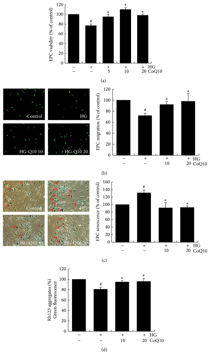
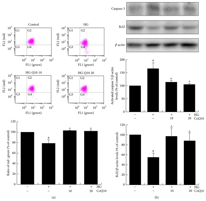
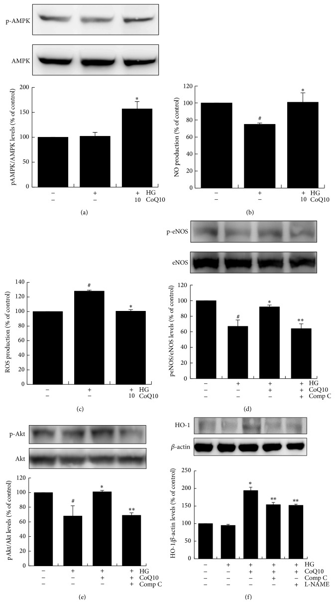
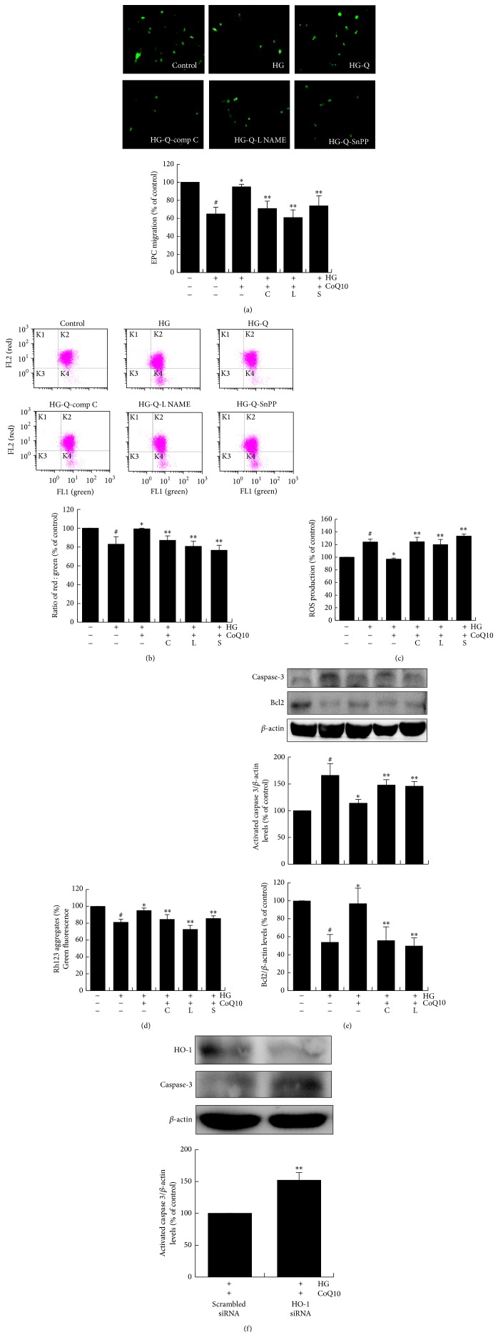
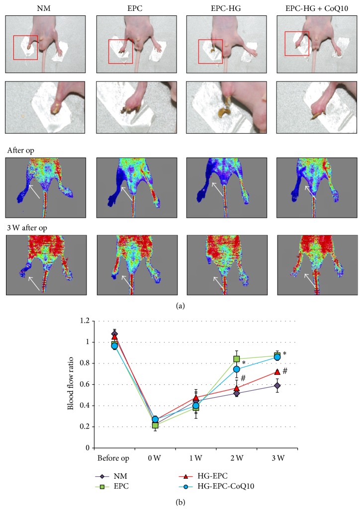
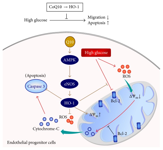
Coenzyme Q10 (CoQ10), an antiapoptosis enzyme, is stored in the mitochondria of cells. We investigated whether CoQ10 can attenuate high glucose-induced endothelial progenitor cell (EPC) apoptosis and clarified its mechanism. EPCs were incubated with normal glucose (5 mM) or high glucose (25 mM) environment for 3 days, followed by treatment with CoQ10 (10 μM) for 24 hr. Cell proliferation, nitric oxide (NO) production, and JC-1 assay were examined. The specific signal pathways of AMP-activated protein kinase (AMPK), eNOS/Akt, and heme oxygenase-1 (HO-1) were also assessed. High glucose reduced EPC functional activities, including proliferation and migration. Additionally, Akt/eNOS activity and NO production were downregulated in high glucose-stimulated EPCs. Administration of CoQ10 ameliorated high glucose-induced EPC apoptosis, including downregulation of caspase 3, upregulation of Bcl-2, and increase in mitochondrial membrane potential. Furthermore, treatment with CoQ10 reduced reactive oxygen species, enhanced eNOS/Akt activity, and increased HO-1 expression in high glucose-treated EPCs. These effects were negated by administration of AMPK inhibitor. Transplantation of CoQ10-treated EPCs under high glucose conditions into ischemic hindlimbs improved blood flow recovery. CoQ10 reduced high glucose-induced EPC apoptosis and dysfunction through upregulation of eNOS, HO-1 through the AMPK pathway. Our findings provide a potential treatment strategy targeting dysfunctional EPC in diabetic patients.


Figures

Figure 1
Figure 1
Morphology and characterization of EPCs from peripheral blood. (a) Peripheral blood mononuclear cells (MNCs) were plated on a fibronectin-coated culture dish on the fourteenth day. EPCs were also characterized by immunofluorescence staining for the expression of (b) DiI-AcLDL, (c) lectin, (d) CD133, (e) kinase insert domain receptor (KDR), (f) platelet/endothelial cell adhesion molecule-1 (CD31), (g) CD34, (h) VE-cadherin, and (i) Von Willebrand factor (vWF). Cells were counterstained with 4′,6-diamidino-2-phenylindole (DAPI) for the nuclei (blue).
Figure 2
Figure 2
Effects of CoQ10 on EPC viability, migration, senescence, and mitochondrial function under high glucose conditions. Cells were cultured with glucose (25 mM) for 3 days, followed by treatment with CoQ10 (5 μM, 10 μM, and 20 μM) for 24 hr. (a) EPC viability was analyzed by MTT assay. (b) A Boyden chamber assay was used with SDF-1 as chemoattractive factor for EPC migration. The migrated cells were stained with fluorescein isothiocyanate UEA-1 (lectin) (green) and counted under the fluorescence microscope. (c) EPC senescence was analyzed by senescence-associated acidic-β-galactosidase activity assay. (d) Cell mitochondrial function was measured by staining with rhodamine 123- (Rh123-) derived green fluorescence 5 mM for 20 min. Data are mean ± SE; n = 6; # P < 0.05 versus control (5 mM glucose); P < 0.05 versus high glucose (HG).
Figure 3
Figure 3
Effect of CoQ10 on EPC apoptosis under high glucose conditions. EPCs were incubated with CoQ10 (10 μM) for 24 hrs in high glucose medium. (a) Mitochondrial apoptosis was detected by JC-1 assay by flow cytometry. Loss of mitochondrial membrane potential (ΔΨm) was performed by the change in JC-1-derived fluorescence from red to green. The ratio of red/green fluorescence represented ΔΨm in EPCs. (b) Expression of activated-caspase 3 and Bcl-2 protein levels were assessed by western blot analysis. Data are mean ± SE; n = 4; # P < 0.05 versus control (5 mM glucose); P < 0.05 versus high glucose (HG).
Figure 4
Figure 4
CoQ10 attenuates ROS and activates NO and AMPK pathway of EPCs under high glucose conditions. EPCs were incubated with CoQ10 (10 μM) under high glucose conditions. (a) AMPK protein phosphorylation levels of EPCs were analyzed by western blot. Data are mean ± SE; n = 6. (b) NO production of EPCs was assessed by staining with NO fluorescent indicator 3-amino,4-aminomethyl-2′,7′-difluorofluorescein (DAF-FM) diacetate (10 μM) for 30 min. (c) ROS production of EPCs was assessed by staining with DCFH-DA (10 μM) for 20 min. The fluorescence intensity was measured using a fluorescent microplate reader. ((d), (e), and (f)) Expressions of Akt protein phosphorylation, eNOS protein phosphorylation, and HO-1 protein were assessed by western blot analysis. Comp C: component C, AMPK inhibitor; L-NAME, NO inhibitor. Data are mean ± SE; n = 4; # P < 0.05 versus control (5 mM glucose); P < 0.05 versus high glucose (HG); ∗∗ P < 0.05 versus HG-CoQ10.
Figure 5
Figure 5
CoQ10 improves high glucose-induced EPCs dysfunction by upregulation of eNOS and HO-1. Cells were cultured with comp C (20 μM), L-NAMEA (100 μM), and SnPP IX (10 μM) for 60 min before CoQ10 incubation under high glucose conditions. (a) EPC migration was measured by Boyden chamber assay. The migrated cells were stained with fluorescein isothiocyanate UEA-1 (lectin) (green) and counted under the fluorescence microscope. (b) Mitochondrial apoptosis was detected by JC-1. Loss of mitochondrial membrane potential (ΔΨm) was assessed by the change in JC-1-derived fluorescence from red to green. The ratio of red/green fluorescence represented ΔΨm in EPCs. (c) ROS production of EPCs was assessed by staining with DCFH-DA. (d) The mitochondrial function was measured using Rh123 dye (5 mM), and the fluorescence intensity was measured at 485-nm excitation and 530-nm emission using a fluorescent microplate reader. (e) Expressions of activated-caspase 3 and Bcl2 protein were assessed by western blot. (f) Cells were transfected with scramble and HO-1 siRNA (10 nM), respectively, in CoQ10-treated EPCs under high glucose conditions, and the expressions of activated-caspase 3 and HO-1 protein were detected by western blot. Comp C: component C, AMPK inhibitor; L-NAME, NO inhibitor; Snpp IX, HO-1 inhibitor. Data are mean ± SE; n = 4; # P < 0.05 versus control (5 mM glucose); P < 0.05 versus high glucose (HG); ∗∗ P < 0.05 versus HG-CoQ10.
Figure 6
Figure 6
Effect of CoQ10-treated EPCs in high glucose medium transplantation on hindlimb perfusion. (a) Serial laser Doppler analyses of hindlimb perfusion revealed before and 3 weeks after hindlimb ischemia surgery in nude mice, which received a transplant with normal saline, EPCs, high glucose-treated EPCs, and high glucose-treated EPC incubated with CoQ10. Low or no perfusion is displayed as blue, whereas the highest perfusion is displayed as red. Arrows indicate ischemic (right) limb after hindlimb ischemia surgery. (b) Quantification analysis of perfusion recovery by laser Doppler perfusion imaging rations (ischemic/normal hindlimb) over time in the different groups. Results are mean ± SE; n = 4; # P < 0.05 versus control (5 mM glucose); P < 0.05 versus high glucose (HG).
Figure 7
Figure 7
The schematic diagram summarizes possible mechanisms by which CoQ10 reduces hyperglycemia-induced endothelial progenitor cell damage. In high glucose condition, CoQ10 improved EPCs migration and apoptosis by ROS and caspase 3 downregulation and mitochondrial membrane potential (ΔΨm) and Bcl-2 upregulation through HO-1 pathway.

References

    1. Federman D. G., Bravata D. M., Kirsner R. S. Peripheral arterial disease: a systemic disease extending beyond the affected extremity. Geriatrics. 2004;59(4):29–32. - PubMed
    1. Adeghate E. Molecular and cellular basis of the aetiology and management of diabetic cardiomyopathy: a short review. Molecular and Cellular Biochemistry. 2004;261(1):187–191. doi: 10.1023/b:mcbi.0000028755.86521.11. - DOI - PubMed
    1. Asahara T., Murohara T., Sullivan A., et al. Isolation of putative progenitor endothelial cells for angiogenesis. Science. 1997;275(5302):964–967. doi: 10.1126/science.275.5302.964. - DOI - PubMed
    1. Rehman J., Li J., Orschell C. M., March K. L. Peripheral blood “endothelial progenitor cells” are derived from monocyte/macrophages and secrete angiogenic growth factors. Circulation. 2003;107(8):1164–1169. doi: 10.1161/01.cir.0000058702.69484.a0. - DOI - PubMed
    1. Vasa M., Fichtlscherer S., Aicher A., et al. Number and migratory activity of circulating endothelial progenitor cells inversely correlate with risk factors for coronary artery disease. Circulation Research. 2001;89(1):e1–e7. - PubMed

Publication types

MeSH terms

LinkOut - more resources