. 2016 Apr 1;594(7):1821-40.
doi: 10.1113/JP271690. Epub 2016 Feb 2.

Kainate receptor pore-forming and auxiliary subunits regulate channel block by a novel mechanism

Affiliations

Kainate receptor pore-forming and auxiliary subunits regulate channel block by a novel mechanism

Patricia M G E Brown et al. J Physiol. .

Abstract

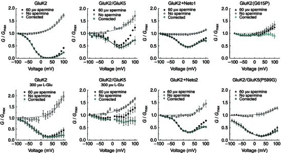
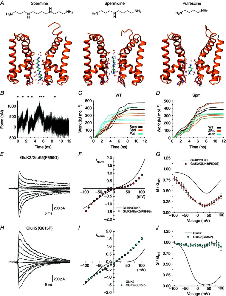
Key points: Kainate receptor heteromerization and auxiliary subunits, Neto1 and Neto2, attenuate polyamine ion-channel block by facilitating blocker permeation. Relief of polyamine block in GluK2/GluK5 heteromers results from a key proline residue that produces architectural changes in the channel pore α-helical region. Auxiliary subunits exert an additive effect to heteromerization, and thus relief of polyamine block is due to a different mechanism. Our findings have broad implications for work on polyamine block of other cation-selective ion channels.

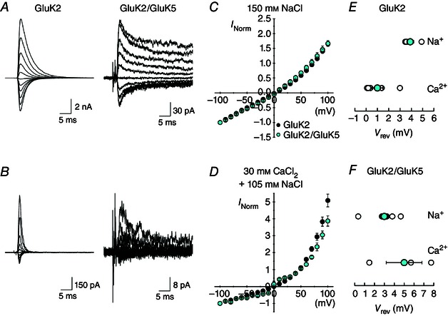
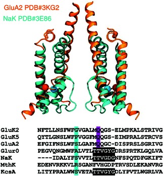
Abstract: Channel block and permeation by cytoplasmic polyamines is a common feature of many cation-selective ion channels. Although the channel block mechanism has been studied extensively, polyamine permeation has been considered less significant as it occurs at extreme positive membrane potentials. Here, we show that kainate receptor (KAR) heteromerization and association with auxiliary proteins, Neto1 and Neto2, attenuate polyamine block by enhancing blocker permeation. Consequently, polyamine permeation and unblock occur at more negative and physiologically relevant membrane potentials. In GluK2/GluK5 heteromers, enhanced permeation is due to a single proline residue in GluK5 that alters the dynamics of the α-helical region of the selectivity filter. The effect of auxiliary proteins is additive, and therefore the structural basis of polyamine permeation and unblock is through a different mechanism. As native receptors are thought to assemble as heteromers in complex with auxiliary proteins, our data identify an unappreciated impact of polyamine permeation in shaping the signalling properties of neuronal KARs and point to a structural mechanism that may be shared amongst other cation-selective ion channels.


Figures

Figure 1
Figure 1. Heteromerization and auxiliary proteins reduce inward rectification of KARs
A, C, E, F, typical membrane currents evoked by 1 mm l‐Glu (1 ms application) at various holding potentials (−100 to +100 mV, 20 mV increments) in the presence of 60 μm internal Spm. Patch numbers: GluK2, 120403p1; GluK2/GluK5, 120417p3; GluK2+Neto1, 130506p3; and GluK2+Neto2, 131104p3. B, D, G, average I–V plots for GluK2 (n = 5), GluK2/GluK5 (n = 4), GluK2+Neto1 (n = 12) and GluK2+Neto2 (n = 7). Data are presented as mean ± SEM. Current values are normalized to the current at −100 mV. H, to obtain the rectification ratio, the peak current at +80 mV was divided by that at −80 mV. Rectification ratios for individual patches are shown as empty circles; columns represent the mean and error bars indicate SEM. One‐way ANOVA, F 3,24 = 60.819, P = 2.32E‐11, post hoc Tukey HSD pairwise comparisons: ****P < 1.33E‐8; n.s., not significant.
Figure 2
Figure 2. Association of GluK2 with GluK5 or auxiliary proteins reduces polyamine block by increasing permeation rates
A, conductance–voltage (G–V) plots for GluK2/GluK5 in the presence (black circles) and absence (white circles) of 60 μm internal Spm. Cyan circles represent the corrected G–V (n = 4), obtained by dividing the Spm G–V by the fit of the Spm‐free G–V. B, corrected G–V plot for GluK2 (n = 5). C, rates of onset, unbinding and permeation for GluK2 (see Methods). k on was estimated using values from Bowie et al. (1998). D, corrected G–V plots for GluK2+Neto1 (cyan, n = 12) and GluK2+Neto2 (orange, n = 7). All data are represented as the mean ± SEM. G–V plots were fit with eqn (2). E and F, comparison of the estimated unbinding (k off, E) and permeation (k perm, F) rates for GluK2 (1), GluK2/GluK5 (2), GluK2+Neto1 (3) and GluK2+Neto2 (4).
Figure 3
Figure 3. G–V corrections for intrinsic outward rectification in the absence of internal polyamines
G–V relationships in the absence (white symbols) or presence (black symbols) of internal polyamines were plotted. The corrected G–V relationships (cyan symbols) were obtained by dividing the value of the spermine G–V by the non‐spermine G–V (G corr = G Spm/G noSpm). Note the importance of performing this correction for each receptor type, as the intrinsic outward rectification properties differ.
Figure 4
Figure 4. Spermine permeability is increased in GluK2/GluK5 and GluK2+Neto2
Electrophysiological traces of GluK2 (A and B, patch 130730p2), GluK2/GluK5 (D and E, patches 121122p1 and 131206p2) and GluK2+Neto2 (G and H, patches 131017p3 and 140410p9) evoked by 1 mm l‐Glu (250 ms) at various holding potentials (−100 to +100 mV, 40 mV increments) in 150 mm NaCl external solution (left) or 90 mm Spm external solution (right). Insets show the current traces at −100 mV in 90 mm Spm. I–V plots in 90 mm external Spm for GluK2 (C, n = 4), GluK2/GluK5 (F, n = 3) and GluK2+Neto2 (I, n = 5). Arrows represent the reversal potentials for GluK2/GluK5 (V rev = −43.2 mV) and GluK2+Neto2 (V rev = −33.6 mV). Insets show the I–V plots ranging from −100 to +100 mV. Grey lines are the fits of the same receptor in the presence of 150 mm external NaCl. Data are represented as means ± SEM. Current values (both Na+ and Spm currents) are normalized to the Na+ current at −100 mV in the same patch.
Figure 5
Figure 5. Ca2+ permeability is similar for GluK2 and GluK2/GluK5 kainate receptors
A, example membrane currents for GluK2 (left, patch 120507p1) and GluK2/GluK5 (right, patch 120522p2) evoked by 1 ms l‐Glu applications (1 mm, in 150 mm NaCl) at various holding potentials (−100 to +100 mV, 20 mV increments) in the absence of internal polyamines. B, example membrane currents for GluK2 (left, same patch as in A) and GluK2/GluK5 (right, same patch as in A) evoked by 1 ms l‐Glu applications (1 mm, in 30 mm CaCl2) at various holding potentials (−100 to +100 mV, 20 mV increments) in the absence of internal polyamines. C, I–V relationships for GluK2 (black, n = 4) and GluK2/GluK5 (cyan, n = 8) in 150 mm NaCl external solution. D, I–V relationships for GluK2 (black, n = 5) and GluK2/GluK5 (cyan, n = 3) in 30 mm CaCl2 external solution. Current values are normalized to the current at −100 mV. E and F, reversal potentials in 150 mm NaCl and 30 mm CaCl2 external solutions for GluK2 (E) and GluK2/GluK5 (F). All data are represented as mean ± SEM.
Figure 6
Figure 6. Prolines in GluK5 pore are predicted to alter pore dimensions
A, sequence alignment of the kainate receptor subunits. A proline is present in the secondary subunits where a conserved glycine is present in primary subunits. This residue is located within a region with a predicted α‐helical structure of the P‐loop. B, crystal structure of the NaK channel (PDB#3E86). Proline residues were mutated in the equivalent α‐helical structure of subunits A and C (yellow spheres). C, side view of the A/C (cyan) and B/D (orange) subunits of the inverted NaK pore before (grey) and after (coloured) 257 ns simulations. D, cross‐pore distance measurements (see black spheres in C) were measured for 500 ns (two repeats for each condition).
Figure 7
Figure 7. The NaK structure is similar to the GluA2 structure
The inverted structure of the open NaK channel (PDB #3E86, cyan) is overlaid with that of the closed GluA2 (PDB #3KG2, orange), illustrating that the two channels share a similar architecture. The proline position is indicated by a sphere. Only two chains are shown and for GluA2, only transmembrane helices M1–3 are included. The two structures are aligned using M2, Cα atoms of residues 573–586 in GluA2 and of residues 50–63 in the NaK channel. The position equivalent to the proline in GluK5 is highlighted by showing the Cα atom of this residue with a sphere (Ser580 in GluA2, Ser57 in NaK). A sequence alignment is shown, highlighting the Gly and Pro residues in GluK2 and GluK5, respectively, which correspond to an Ser in NaK.
Figure 8
Figure 8. RMSD is larger in helices containing prolines
The RMSD values calculated for the Cα atoms of the P‐loop helix (residues 50–62) of each chain of the NaK channel over the course of the 500 ns simulation. The alignment and RMSD calculation were performed for one chain at a time, aligning by residues 50–62 of the given helix for which the RMSD is calculated. WT helices are shown in black/grey, helices from the 2Pro simulation in red/orange and those of the 4Pro simulation in blue/cyan.
Figure 9
Figure 9. Proline in the M2 helix controls spermine block and permeation
A, each of the three polyamines binding in the NaK filter region, from left to right: spermine (Spm), spermidine (Spd), putrescine (Put). For simplicity, only chains A and C of the protein are included and non‐polar hydrogen atoms of the protein are omitted. Carbon atoms of the ligands are shown in cyan. B, an example of a force profile illustrating the force added when pulling Spm out of the WT filter towards the intracellular side. C, work profiles for pulling the three different polyamines to the intracellular side in the WT protein. Spm results are shown in black, Spd in orange and Put in cyan. The work involved in Spm release is generally larger than for release of the smaller polyamines. D, work profiles for pulling Spm to the intracellular side for the WT protein (black), the 2Pro mutant (orange) and the 4Pro mutant (cyan). E and H, example responses of GluK2/GluK5(P599G) (E, patch 130610p7) and GluK2(G615P) (H, patch 130606p2) at various holding potentials (−100 to +100 mV, 20 mV increments) in the presence of 60 μm internal spermine. Average I–V plots (F, I) and corrected G–V plots (G, J) for these receptors in the presence of internal spermine. Relationships for GluK2 and GluK2/GluK5 (grey lines in G and J) are shown for comparison. Data are represented as mean ± SEM. Current values are normalized to the current at −100 mV.
Figure 10
Figure 10. Snapshots illustrating the interactions as Spm is pulled toward the intracellular side of the membrane
Snapshots corresponding to the asterisks in Fig. 9 B. Spermine is shown with cyan carbon atoms. Hydrogen bonds between spermine and the protein [dist(H–O) < 2.5 Å] are indicated with black lines.
Figure 11
Figure 11. G615P mutation and Neto2 association increase spermine permeation of GluK2
Example responses of GluK2 (A, same data as in Fig. 3, GluK2(G615P) (B, patch 130906p2) and GluK2(G615P)+Neto2 (C, patch 140814p4) in 90 mm external Spm, at various holding potentials (−100 to +100 mV, 40 mV increments). Insets show the response in 90 mm external Spm at −100 mV. Average I–V plot for GluK2 (D, same data as in Fig. 3), GluK2(G615P) (E, n = 5) and GluK2(G615P)+Neto2 (F, n = 4) in 90 mm external Spm; arrows indicate reversal potentials for GluK2(G615P) (V rev = −25.5 mV) and GluK2(G615P)+Neto2 (V rev = −15.6 mV). Insets show the I–V plots ranging from −100 to +100 mV. Grey lines are the fits of the same receptor in the presence of 150 mm external NaCl. Data are represented as means and SEM. Current values (both Na+ and Spm currents) are normalized to the Na+ current at −100 mV in the same patch. G, left: summary plot showing the V rev (in 90 mm Spm) for the various receptors tested. Right: summary plot showing the calculated relative Spm permeabilities (P Spm/P Na). One‐way ANOVA, F 3,12 = 9.801, P = 0.002; post hoc Tukey HSD pairwise comparisons: *P = 0.027 and **P = 0.005 and 0.002 for G615P+Neto2–GluK2+Neto2 and G5615P+Neto2–GluK2/GluK5 comparisons, respectively.

References

    1. Aizenman CD, Munoz‐Elias G & Cline HT (2002). Visually driven modulation of glutamatergic synaptic transmission is mediated by the regulation of intracellular polyamines. Neuron 34, 623–634. - PubMed
    1. Alam A & Jiang Y (2009. a). High‐resolution structure of the open NaK channel. Nat Struct Mol Biol 16, 30–34. - PMC - PubMed
    1. Alam A & Jiang Y (2009. b). Structural analysis of ion selectivity in the NaK channel. Nat Struct Mol Biol 16, 35–41. - PMC - PubMed
    1. Bahring R, Bowie D, Benveniste M & Mayer ML (1997). Permeation and block of rat GluR6 glutamate receptor channels by internal and external polyamines. J Physiol 502, 575–589. - PMC - PubMed
    1. Barberis A, Sachidhanandam S & Mulle C (2008). GluR6/KA2 kainate receptors mediate slow‐deactivating currents. J Neurosci 28, 6402–6406. - PMC - PubMed

Publication types